
Practical Guide: A Practical Guide to Building a Graphene Battery Safety Sensor
Build a flexible graphene battery sensor for real-time temperature and strain monitoring, a crucial step toward preventing thermal runaway in Li-ion cells.
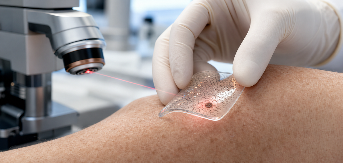
Graphene skin patch research is moving from simple wearable sensors toward active therapeutic materials. A 2026 ACS Nano paper by Xiaoyu Xu, Li Cheng, Bohan Li, and colleagues reports one of the more striking examples: a stretchable, transparent, laser-induced graphene patch designed to sit on the skin, remain inactive under normal conditions, and release copper ions only when gently warmed by light.
The target was melanoma, the most dangerous form of skin cancer and a disease where localized treatment is still difficult. Traditional surgical excision remains the standard approach for many early lesions, while advanced disease may require immunotherapy, targeted therapy, radiation, or other systemic treatment. The new patch is not a clinical product and should not be interpreted as a substitute for medical care. It has been tested in cells and in a preclinical mouse model, not in humans.
Still, the study is important because it shows how graphene can act as more than a passive support material. In this device, laser-induced graphene provides a porous, photothermally active structure. Copper(II) oxide nanoparticles provide the therapeutic copper source. A soft PDMS silicone matrix gives the patch flexibility, transparency, and skin contact. When these pieces are combined, the result is a material system that can deliver a controlled local chemical effect under mild heating.
The patch described in the ACS Nano paper is a soft LIG-Cu/PDMS hybrid. LIG stands for laser-induced graphene, a porous carbon material typically made by using a laser to convert a carbon-rich precursor into graphene-like conductive networks. This approach is attractive because it can pattern conductive graphene structures directly and can create a high-surface-area framework without requiring a separate high-temperature graphene transfer process.
In this study, the active component was copper(II) oxide embedded into laser-induced graphene. The LIG-Cu structure was then incorporated into PDMS, a flexible silicone widely used in biomedical and wearable-device research because it is soft, transparent, chemically stable, and able to conform to curved surfaces.
That mechanical design matters. Skin is not flat, rigid, or still. A tumor patch needs to bend with the body, stay in close contact with tissue, allow some breathability, and avoid causing unnecessary irritation. The researchers emphasized that the patch was stretchable, transparent, reusable, and easy to apply. Those features are not cosmetic details; they are part of why skin-mounted therapeutic devices are so challenging to engineer.
The material also had to remain relatively inert before activation. A patch that releases copper constantly would be much harder to control. The key idea in this paper is a heat-and-release mechanism: under ordinary conditions, the device is stable; under mild photothermal stimulation, it releases copper ions locally into the tumor area beneath it.
Laser-induced graphene is well suited to this kind of patch because it can convert light into heat efficiently. When a low-power laser or simulated light source activates the patch, the graphene network absorbs the energy and raises the local temperature. In the reported tests, the target temperature was about 42 C, or 108 F.
That temperature is important. It is warm enough to stimulate copper ion release from the CuO-loaded porous graphene, but it is far below the kind of aggressive thermal ablation used to burn tissue. In other words, the patch is not mainly trying to cook the tumor. It is using mild heat as a trigger for localized ion release and as part of a broader photothermal-chemical treatment strategy.
Graphene's porous structure also helps. The pores can hold active materials, expose surface area, and support interaction with the surrounding matrix. In a therapeutic patch, surface area and transport pathways are crucial because the device has to deliver active ions into a biological microenvironment without simply dumping material everywhere.
The result is a smart materials platform: graphene absorbs light, graphene helps host copper oxide, the PDMS matrix keeps the device soft and wearable, and the heat stimulus controls when copper ions are released.
Copper is essential in biology, but excess copper can become toxic to cells. Cancer researchers are increasingly interested in copper because tumor cells often have altered metal metabolism, oxidative stress balance, mitochondrial activity, and redox chemistry. If copper can be delivered locally and selectively, it may push cancer cells past their stress threshold while limiting systemic side effects.
The ACS Nano study reports that, after activation, the patch released Cu2+ ions into melanoma cells directly beneath the treated area. Those ions increased reactive oxygen species, damaged mitochondria, and triggered several forms of programmed cell death.
The paper highlights apoptosis, cuproptosis, and ferroptosis. Apoptosis is the best-known programmed cell death pathway, often described as controlled cellular self-destruction. Cuproptosis is a newer copper-dependent death mechanism associated with mitochondrial metabolism and copper binding to lipoylated proteins. Ferroptosis is an iron-associated cell death pathway tied to lipid peroxidation and oxidative damage.
Why does it matter that multiple pathways are involved? Cancer cells can resist treatment by adapting around a single mechanism. A local therapy that activates several stress and death routes at once may reduce the chance that melanoma cells simply survive, migrate, or recover after partial damage. That does not prove clinical success, but it makes the mechanism more interesting than simple heat treatment alone.
The most attention-grabbing number from the study is the reported 97 percent reduction in melanoma lesions within 10 days in a mouse model. According to ACS-distributed summaries of the work, the mice received two one-hour phototherapy sessions, one on day 1 and one on day 5. The patch was activated with a low-power laser, raising the local temperature to about 42 C and releasing copper ions into the tumor region.
By day 10, treated lesions were dramatically reduced compared with the untreated controls. Tissue analysis also suggested that cancer cells did not migrate beyond the tumor border in the treated animals. That second point matters because melanoma is dangerous not only because the primary lesion grows, but because it can invade and metastasize.
The researchers also reported no significant damage to surrounding healthy skin and no harmful copper accumulation in major organs or blood. That is one of the most important safety signals in the study. Copper can be therapeutically useful only if it can be kept where it is needed. A patch that shrinks a lesion but spreads toxic copper systemically would not be a good trade.
Preclinical mouse data should always be handled carefully. Mice are not humans, implanted or induced tumors are not the same as naturally evolving human melanoma, and a 10-day study cannot answer long-term recurrence, immune memory, dosing, skin-type variability, or clinical safety questions. But as a proof of concept, the magnitude of the local response is notable.
One of the stronger parts of the concept is that the patch may do more than kill cells directly under the device. The paper reports that treatment stimulated antitumor immune activity and helped suppress invasion and metastasis-related behavior.
This is plausible because when tumor cells die under oxidative and mitochondrial stress, they can release tumor-associated antigens and danger signals. Those signals may help the immune system recognize tumor material and respond beyond the immediate treated area. In cancer therapy, this is a major goal: local tumor destruction that also educates or activates systemic immunity.
However, this is also where caution is needed. Immune effects in mouse models can be real and still fail to translate cleanly into humans. Human melanoma exists inside a complex immune environment shaped by patient genetics, tumor mutations, prior treatment, age, sun exposure, and many other variables. The patch's immune contribution will need much deeper study before anyone can claim durable metastasis prevention in patients.
For materials scientists, the interesting point is that the device is not just a drug delivery patch. It is a material-triggered microenvironment intervention. The patch changes local copper chemistry, oxidative stress, mitochondrial function, cell death signaling, and possibly immune presentation. That is a high bar for a thin flexible material.
Graphene is often discussed in broad terms: stronger composites, better batteries, faster electronics, more sensitive sensors. This paper shows another direction: graphene as a controllable interface between energy, chemistry, and living tissue.
The patch uses several graphene-relevant advantages at the same time. It needs photothermal conversion, because light must be turned into controlled heat. It needs porosity, because copper oxide has to be embedded and released in a regulated way. It needs mechanical compliance, because the device must conform to skin. It needs chemical stability, because the patch should remain inactive and safe until triggered.
That combination is hard to get from a conventional flat film. Laser-induced graphene gives researchers a way to build patterned, porous, conductive, light-responsive structures directly into flexible device formats. For biomedical use, that opens possibilities beyond melanoma: wound treatment, antibacterial patches, localized drug release, wearable diagnostics, and combined sensor-therapy systems.
The strongest commercial lesson is not that every skin cancer patch will use graphene. The lesson is that graphene-based structures can be engineered as active materials platforms, not only as additives. They can host nanoparticles, respond to light, manage heat, and interface with soft tissue in one integrated system.
The reported patch has several practical attributes that make it easy to understand why researchers are interested in it. It is transparent, so clinicians could potentially see the treated area. It is stretchable, so it can remain in contact with moving skin. It is reusable in the reported experimental setting, which could reduce material waste and cost. It is activated externally, so treatment timing can be controlled.
For patients, a future version of such a patch would be far less invasive than surgical excision if it proved safe and effective. It could also be attractive for lesions in sensitive locations where tissue preservation matters. For clinicians, a patch-based treatment could offer a standardized local therapy with clear dosing parameters: patch area, light intensity, temperature, treatment time, and number of sessions.
But there are also practical questions. Human lesions vary in thickness, pigmentation, shape, vascularity, and depth. Melanoma diagnosis depends on pathology. Many suspicious lesions need biopsy, not just treatment. A patch would need to fit into that clinical workflow. It would also need strict temperature control, clear margins, reliable copper release kinetics, and strong evidence that residual malignant cells are not left behind.
The device is promising because it is convenient. It is not proven simply because it is convenient.
Before any human use, several hard questions remain.
First, researchers need longer safety studies. Copper accumulation in major organs was not significant in the reported mouse study, but long-term local tissue effects, repeated exposure, and different dosing schedules require more work.
Second, the treatment needs testing in more realistic tumor models. Human melanoma can be heterogeneous, invasive, and resistant. A patch has to work across lesion geometries and depths, not only in a controlled preclinical setup.
Third, researchers must define treatment margins. In surgery, clinicians remove tissue around the visible lesion to reduce the risk of leaving cancer cells behind. A noninvasive patch would need an equivalent logic: how large should the patch be, how far beyond the visible lesion should treatment extend, and how should complete response be confirmed?
Fourth, the system must be manufactured reproducibly. Laser-induced graphene can vary depending on precursor, laser parameters, atmosphere, and post-processing. Copper oxide loading, PDMS bonding, transparency, flexibility, ion release rate, and photothermal response all need process control.
Finally, regulatory and clinical pathways will be demanding because this is a combination of material, device, light activation, and local therapeutic ion release. That does not make translation impossible, but it does mean the journey from ACS Nano paper to dermatology clinic will be long.
The ACS Nano study on a photothermally stimulated laser-induced graphene melanoma patch is a strong example of where advanced materials are heading. The patch combines LIG, copper(II) oxide nanoparticles, and soft PDMS into a transparent device that remains quiet until activated by mild heat. In preclinical mouse tests, two one-hour treatments reduced melanoma lesions by 97 percent within 10 days while sparing surrounding skin and avoiding obvious systemic copper accumulation.
The most important point is not that a skin cancer cure has arrived. It has not. This is early experimental work and needs substantial validation before clinical use. The important point is that graphene can be engineered into active, responsive biomedical systems that control when and where therapy is delivered.
For USA Graphene readers, this is the broader signal: graphene's value in medicine may come from integration. When graphene's photothermal behavior, porosity, conductivity, flexibility, and compatibility with soft polymers are combined with therapeutic chemistry, the material becomes part of the treatment mechanism itself.
Source: Xiaoyu Xu, Li Cheng, Bohan Li, et al. "A Stretchable, Transparent, Photothermally Stimulated Laser-Induced Graphene Patch for Noninvasive Skin Tumor Treatment." ACS Nano, 2026. DOI: 10.1021/acsnano.5c21102.
Serious about B2B integration? Test our premium Pulsed Electrical Resistive Carbon Heating turbostratic graphene in your lab. 100g sample packs available now.
Explore More
Build a flexible graphene battery sensor for real-time temperature and strain monitoring, a crucial step toward preventing thermal runaway in Li-ion cells.

Research on 30° quasi-periodic twisted bilayer graphene reveals a unique, fractal-like energy structure under magnetic fields, paving the way for novel...

Researchers discover naturally formed graphene sheets and carbon ovals in stingless bee hives, creating a novel emergent material with powerful blue light...