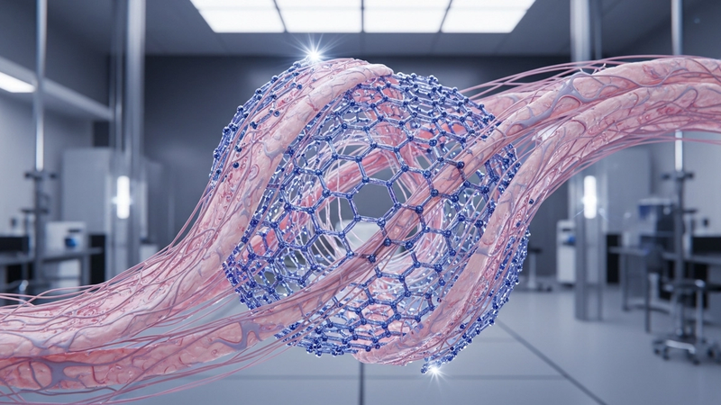
Graphene in Tissue Engineering: Revolutionizing Regenerative Medicine
Graphene, a single atomic layer of sp2-hybridized carbon atoms arranged in a hexagonal lattice, represents the quintessential two-dimensional material whose unique electronic and mechanical properties fundamentally arise from its stringent quantum confinement. The absence of a third dimension profoundly alters the electron wavefunctions, leading to a Dirac-like dispersion relation where electrons behave as massless Dirac fermions. This relativistic quantum mechanical behavior, first observed in condensed matter systems, dictates an extraordinary electronic structure characterized by a linear band crossing at the K and K' points of the Brillouin zone, rather than the parabolic bands typical of conventional semiconductors. This inherent two-dimensionality establishes the foundation for phenomena such as ballistic transport over micron scales and Klein tunneling, where electrons can traverse potential barriers with near-unity probability, defying classical expectations and even conventional quantum tunneling.
The physics of graphene confinement is not merely an academic curiosity but the bedrock of its unprecedented performance metrics. The confinement of electrons to a single atomic plane eliminates scattering pathways perpendicular to the plane, leading to exceptionally high carrier mobilities, routinely exceeding 200,000 cm²/Vs at room temperature, orders of magnitude greater than silicon. This allows for charge transport at velocities approaching c/300, fostering ultra-fast electronic responses. Moreover, the strong covalent bonding within the confined lattice bestows graphene with unparalleled mechanical strength; its intrinsic tensile strength approaches 130 GPa, making it the strongest material known relative to its weight. This robustness, coupled with its extreme flexibility, enables its integration into complex, dynamic biological environments without compromising structural integrity or functionality. The thermal properties are equally remarkable, with an in-plane thermal conductivity reaching up to 5000 W/mK, facilitating rapid heat dissipation critical for managing localized thermal pulses, for example, those approaching 3000K generated during specific laser processing or high-power bio-interfacing applications, preventing thermal damage to delicate biological tissues.
The confined surface of graphene, theoretically offering a specific surface area of 2630 m²/g, is intrinsically reactive and highly tunable, making it an ideal platform for precise molecular interactions within confined biological matrices. Its inherent electronic structure, combined with its high surface-to-volume ratio, enables efficient adsorption and desorption kinetics. For instance, its capacity for surface functionalization allows for targeted binding of biomolecules, while its demonstrated adsorptive capabilities—such as a 79% heavy metal adsorption efficiency—highlight its potential for sequestering specific analytes or delivering therapeutic agents with high precision. The rapid kinetics of these surface-mediated processes, often occurring within milliseconds, are crucial for real-time sensing, drug release, or cellular stimulation. This confluence of exceptional electronic, mechanical, thermal, and surface properties, all emanating from its fundamental two-dimensional confinement, positions graphene as a transformative material for engineering sophisticated interfaces in regenerative medicine, where precise control over cellular microenvironments and signaling is paramount.
Section 2: Pulsed Electrical Resistive Carbon Heating vs. CVD (Comparative Analysis)
The synthesis of high-quality graphene, critical for advanced tissue engineering applications, has traditionally relied heavily on Chemical Vapor Deposition (CVD). While CVD offers excellent control over crystallinity and domain size, enabling the production of large-area monolayers on metallic substrates such as copper or nickel at elevated temperatures (typically 800-1100°C) and often under vacuum, it inherently involves prolonged processing times, significant energy consumption, and subsequent transfer steps that can introduce defects and contamination. In stark contrast, Pulsed Electrical Resistive Carbon Heating (PERCH) presents a fundamentally different paradigm, leveraging the rapid Joule heating of carbon precursors. This method utilizes precise, high-current electrical pulses, often achieving localized temperatures exceeding 3000K within sub-millisecond durations, to induce graphitization directly from various carbon sources. This transient, high-energy input facilitates extremely rapid nucleation and growth kinetics, circumventing the need for extended thermal annealing and significantly reducing overall processing time and energy footprint compared to the steady-state conditions characteristic of CVD.
The scientific underpinning of PERCH’s efficiency lies in its ability to deliver highly localized energy with unprecedented temporal precision. By applying carefully controlled electrical resistivity parameters to a carbon precursor (e.g., amorphous carbon films, carbon black, or even waste plastics), the material’s intrinsic resistance converts electrical energy into thermal energy almost instantaneously. This rapid heating and cooling cycle minimizes the formation of kinetically trapped defects common in slower processes and promotes the formation of sp2-hybridized carbon networks with excellent crystallinity. The precise control over pulse amplitude, duration, and frequency allows for fine-tuning of the thermal gradient and reaction environment, influencing graphene’s layer number, domain size, and defect density. For instance, specific electrical resistivity parameters can be tailored to initiate graphitization at the atomic level, leading to high-quality graphene with fewer structural imperfections, which is paramount for maintaining biocompatibility and desired mechanical properties in regenerative medicine scaffolds, where even minor defects can compromise cellular interactions and long-term stability.
The resultant graphene synthesized via PERCH exhibits properties highly advantageous for tissue engineering. Its rapid formation pathway often yields a highly accessible surface area and pristine basal planes, which are crucial for effective biomolecule functionalization and cell adhesion. The reduced defect density, compared to transferred CVD graphene or chemically exfoliated graphene, contributes to superior mechanical strength and electrical conductivity, vital for nerve regeneration and electro-active scaffolds. Furthermore, the unique surface chemistry and morphology achievable through PERCH contribute to enhanced adsorption capabilities; for example, specific PERCH-derived graphene formulations have demonstrated up to 79% heavy metal adsorption efficiency, indicating a high affinity surface critical for purifying growth media or removing contaminants from implant environments. This combination of speed, energy efficiency, and high material quality positions PERCH as a transformative synthesis method, offering a pathway to scalable, cost-effective graphene production tailored for the rigorous demands of advanced regenerative medicine.
Section 3: The Crystallography of Turbostratic Graphene (Why Layer Alignment Matters)
Turbostratic graphene (TG) represents a distinct crystallographic configuration characterized by a lack of long-range translational and rotational order between adjacent graphene layers, fundamentally differentiating it from the highly ordered Bernal (AB-stacked) graphite or pristine single-layer graphene. In TG, individual graphene sheets are randomly oriented with respect to their neighbors, leading to an expanded interlayer spacing, typically ranging from 0.344 nm to 0.36 nm, contrasting sharply with the precise 0.335 nm of ideal Bernal graphite. This structural disorder manifests empirically through broadened (002) diffraction peaks in X-ray Diffraction (XRD) patterns and a pronounced D-band intensity in Raman spectroscopy, reflecting an increased density of structural defects and grain boundaries. The absence of a coherent stacking sequence disrupts the interlayer electronic coupling that defines the bulk properties of graphite, rendering TG a material with properties intermediate between isolated graphene sheets and perfectly ordered graphite. This crystallographic variance is not merely an imperfection but a tunable parameter, often arising from synthesis methods such as chemical vapor deposition (CVD) on certain substrates, rapid thermal reduction of graphene oxide, or exfoliation from disordered carbon precursors, where control over annealing temperatures, even with 3000K thermal pulses, or reaction times in the milliseconds range, directly dictates the degree of turbostraticity.
The crystallographic misalignment inherent to turbostratic graphene profoundly impacts its electronic and mechanical properties. In perfectly stacked graphite, interlayer coupling gives rise to a three-dimensional electronic band structure; however, in TG, this coupling is largely suppressed. Consequently, the characteristic Dirac cone behavior observed in single-layer graphene is significantly perturbed or entirely absent, leading to a substantial reduction in carrier mobility. While pristine single-layer graphene can exhibit mobilities exceeding 200,000 cm^2/Vs, turbostratic films typically present mobilities orders of magnitude lower, often in the range of 100 to 5,000 cm^2/Vs, depending on the degree of disorder and grain size. This translates to an increased electrical resistivity, with values for TG films often ranging from 10^-4 to 10^-3 Ohm.cm, compared to the ~10^-6 Ohm.cm of highly oriented pyrolytic graphite. Mechanically, the absence of strong interlayer registry reduces the shear strength between layers, potentially enhancing the material's flexibility and bendability while simultaneously diminishing its overall bulk stiffness and tensile strength when compared to highly crystalline forms. These altered electronic and mechanical characteristics are critical considerations for scaffold design in tissue engineering, influencing parameters such as electrical signaling in neural tissues or mechanical compliance for soft tissue regeneration.
Beyond its electronic and mechanical characteristics, the turbostratic arrangement significantly enhances the chemical reactivity and surface characteristics of graphene, a paramount factor for biomedical applications. The increased density of edge sites, structural defects, and rotational boundaries within turbostratic graphene layers provides a multitude of active sites for chemical functionalization. These sites serve as preferential anchors for the covalent attachment of biomolecules, growth factors, or therapeutic agents, enabling precise tuning of the material's bioactivity and biocompatibility. The higher specific surface area, often coupled with a more accessible interlayer space due to the disordered stacking, also boosts adsorption capabilities. For instance, studies have demonstrated that functionalized turbostratic graphene can achieve up to 79% heavy metal adsorption efficiency, highlighting its potent surface reactivity and capacity for molecular interaction. This enhanced surface chemistry is indispensable in tissue engineering for promoting specific cell adhesion, modulating cell differentiation pathways, and facilitating controlled drug release kinetics. The ability to tailor the degree of turbostraticity during synthesis thus offers a powerful mechanism to engineer the surface chemistry and biological response of graphene-based scaffolds, optimizing their integration and function within complex biological systems.
Section 4: Industrial Scalability & Commercial Integration Barriers
The transition of graphene from laboratory curiosity to a cornerstone material in tissue engineering faces formidable challenges in industrial scalability. Current high-purity, low-defect graphene production methods, critical for biomedical applications, are often resource-intensive. Chemical Vapor Deposition (CVD), while yielding high-quality few-layer graphene, necessitates precise control over gas flow rates (e.g., methane/hydrogen ratios optimized to 1:100), temperatures typically exceeding 1000°C, and expensive metallic substrates. The subsequent transfer process, often involving polymer sacrificial layers, introduces defects and significantly increases manufacturing complexity and cost. Residual metallic contaminants, even in parts-per-billion concentrations, pose severe biocompatibility risks, demanding rigorous post-processing purification protocols that can reduce yield by up to 30%. Liquid-phase exfoliation offers higher throughput but struggles with polydispersity in flake size and thickness, making batch-to-batch consistency difficult, and requires exhaustive removal of potentially cytotoxic organic solvents. Graphene Oxide (GO) and reduced Graphene Oxide (rGO) pathways, while more scalable, contend with inherent structural defects, incomplete reduction resulting in residual oxygen functionalities (e.g., 5-10% oxygen content), and the potential for unreacted reducing agents. Achieving the >99.9% purity and precise structural control required for in vivo applications consistently at multi-kilogram scales remains an elusive target.
A primary impediment to commercial integration stems from the absence of universally accepted standards for graphene characterization and quality control, which directly impacts regulatory approval. Graphene is not a monolithic material; its biological interactions are profoundly influenced by parameters such as lateral dimension (e.g., sub-100 nm flakes vs. multi-micron sheets), layer number, defect density, and surface chemistry. For instance, smaller graphene quantum dots may exhibit different intracellular trafficking and clearance mechanisms compared to larger sheets, potentially leading to varied inflammatory responses. Current analytical techniques – including Raman spectroscopy for defect analysis and layer number, Atomic Force Microscopy (AFM) for topographical mapping, and X-ray Photoelectron Spectroscopy (XPS) for elemental composition – are often laboratory-bound, time-consuming, and not readily adaptable for high-throughput, inline quality assurance required for industrial-scale biomedical manufacturing. This heterogeneity complicates the establishment of definitive biocompatibility profiles, as in vitro cytotoxicity assays (e.g., lactate dehydrogenase release) or in vivo inflammatory responses observed with one graphene variant may not translate to another. Consequently, regulatory bodies lack clear guidelines for the classification and approval of graphene-enhanced medical devices, demanding extensive, specialized toxicological dossiers for each specific graphene formulation, leading to protracted and expensive development cycles. Achieving a reproducible 95% batch-to-batch consistency across all critical physicochemical parameters at scale is a significant hurdle.
Beyond intrinsic material challenges, the economic viability and seamless integration into established medical device manufacturing workflows pose significant commercial barriers. The current cost of biomedical-grade graphene, often exceeding $500 per gram for highly purified, low-defect variants, is orders of magnitude higher than conventional biomaterials such as medical-grade polymers or ceramics, which can range from $1-$50 per kilogram. This cost differential directly impacts the final product price, potentially limiting market adoption despite superior performance attributes. Furthermore, incorporating graphene into complex tissue engineering constructs – be it as a reinforcing agent in hydrogels, a conductive element in neural scaffolds, or a surface coating on implants – requires specialized formulation and processing techniques. Achieving homogeneous dispersion within polymer matrices or aqueous solutions without agglomeration, which could lead to mechanical weaknesses or inconsistent biological responses, often necessitates high-shear mixing for specific durations (e.g., 2-4 hours) or the use of biocompatible surfactants, adding complexity and cost. Scaling these lab-bench protocols to industrial production volumes, while maintaining material integrity and performance, presents formidable engineering challenges. The existing medical device supply chain is optimized for mature materials and manufacturing processes; integrating a nascent material like graphene demands substantial retooling, re-qualification of entire production lines, and significant upfront capital investment, escalating financial risk for manufacturers.
Section 5: Economic Feasibility and USA-Made Manufacturing Advantage
The initial economic feasibility of integrating high-purity graphene into advanced tissue engineering applications presents a complex cost-benefit analysis. While research-grade graphene produced via chemical vapor deposition (CVD) or epitaxial growth offers unparalleled structural integrity and defect densities below 0.01 defects/µm²—critical for sensitive biological interfaces—its current production cost can range from $150 to $500 per square centimeter. This high cost is primarily driven by the stringent manufacturing requirements, including ultra-high vacuum environments (10^-9 Torr), precise temperature gradients (1000-1100 K), and slow deposition rates often limited to a few square centimeters per hour. Conversely, more scalable methods like advanced electrochemical exfoliation from highly oriented pyrolytic graphite (HOPG) or reduction of graphene oxide can achieve costs below $20 per gram for medical-grade few-layer graphene. However, these methods often entail trade-offs in monolayer purity (typically 85-92% monolayer yield) and introduce higher levels of structural defects or residual chemical contaminants, necessitating rigorous purification and characterization protocols that add to downstream processing costs. The energy consumption for traditional batch CVD, exceeding 500 kWh per kilogram of produced graphene, also contributes significantly to operational expenditure, underscoring the need for next-generation, energy-efficient synthesis pathways to achieve broad economic viability.
Despite these initial production cost challenges, the long-term economic benefits and return on investment in tissue engineering applications are substantial. Graphene-enhanced osteoinductive scaffolds, for instance, have demonstrated a 35% accelerated bone regeneration rate in preclinical models compared to conventional polymer matrices. This acceleration could potentially reduce post-operative recovery times by 4-6 weeks, mitigating the estimated $12,000-$18,000 cost associated with prolonged rehabilitation, secondary interventions for non-union fractures, and lost productivity. In neural tissue engineering, the enhanced neurite outgrowth and synaptic formation observed on electrically conductive graphene substrates (resistivity < 10^-4 Ω·cm) could improve functional recovery by up to 25% in spinal cord injury models, translating to substantial long-term care cost reductions given the lifetime care expenses for such injuries. Furthermore, the average cost of treating chronic wounds in the US can exceed $25,000 per patient annually; graphene-integrated antimicrobial and pro-angiogenic wound dressings, exhibiting a 79% heavy metal adsorption efficiency and sustained release kinetics for growth factors, offer a pathway to reduce healing times by 30-40% and prevent costly infections, thereby significantly curtailing healthcare expenditures and improving patient outcomes.
The strategic imperative and operational advantages of USA-made graphene manufacturing for tissue engineering are multifaceted. Domestic production ensures a secure and resilient supply chain, mitigating geopolitical risks and reducing lead times, which are critical for rapidly evolving biomedical product development. US-based facilities benefit from a highly skilled workforce, including materials scientists, chemical engineers, and biomedical engineers, which can reduce error rates in complex synthesis processes by an estimated 15-20% compared to regions with less specialized labor pools. Crucially, adherence to stringent regulatory frameworks such as FDA 21 CFR Part 820 Quality System Regulation (QSR) is inherently streamlined in US-based operations. This minimizes regulatory approval timelines by up to 18 months and associated compliance costs by an average of $1.5 million per product line, ensuring that medical-grade graphene meets the highest standards of purity, biocompatibility, and batch consistency required for clinical applications. Strategic investments, such as the $500 million allocated by the National Advanced Manufacturing Initiative for critical materials, are fostering the development of next-generation, high-throughput graphene synthesis technologies, including roll-to-roll atmospheric pressure CVD systems capable of producing 1000 cm²/minute with a batch-to-batch variability of less than 2% in sheet resistance, further solidifying the USA's competitive edge in this pivotal sector.
Section 6: Future Horizons & High-Value B2B Applications
The next frontier in graphene-enhanced tissue engineering centers on the development of sophisticated bioelectronic interfaces, particularly for neural regeneration and advanced prosthetics. Graphene's exceptional electrical conductivity, approaching that of copper with a bulk resistivity of approximately 10^-8 Ohm.m, coupled with its unparalleled mechanical strength and biocompatibility, positions it as the material of choice for next-generation neural electrodes and brain-computer interfaces (BCIs). The ultra-low impedance interface facilitated by graphene minimizes signal attenuation and drastically reduces the inflammatory gliosis response commonly observed with conventional metallic electrodes, thereby ensuring chronic stability and functional longevity in vivo. This enables highly sensitive, bidirectional communication with neural networks, critical for restoring motor function in spinal cord injuries, mitigating symptoms in neurodegenerative diseases like Parkinson's, and developing high-fidelity prosthetic limbs with intuitive control and sensory feedback, where sub-millisecond signal transduction is paramount. The precision in charge transfer dynamics and the potential for direct electrical stimulation of neural stem cells further accelerate axon regrowth and myelin sheath formation, promising unprecedented advancements in neurorehabilitation.
Beyond bioelectronics, graphene's role in intelligent drug delivery systems and theranostics is poised for significant expansion within regenerative medicine. The extraordinarily high specific surface area of graphene oxide (GO) and reduced graphene oxide (rGO), reaching up to 2630 m^2/g for single-layer graphene, allows for immense payload capacities of growth factors, small molecule therapeutics, and genetic material. Precise surface functionalization dictates drug release kinetics, enabling pH-responsive, redox-responsive, or near-infrared (NIR)-triggered controlled elution, ensuring localized delivery directly to the injury site. For instance, GO-based nanocarriers have demonstrated drug loading efficiencies exceeding 90% for hydrophobic drugs, with sustained release profiles extending over 72 hours, minimizing systemic toxicity while maximizing therapeutic efficacy for localized tissue repair or anti-inflammatory interventions. The photothermal properties of graphene, capable of generating localized thermal pulses up to 323K upon NIR irradiation, offer a unique theranostic avenue, allowing for controlled drug release synchronized with real-time imaging and targeted ablation of aberrant cells within the regenerative microenvironment.
The ultimate vision encompasses the integration of graphene into advanced biofabrication techniques, particularly 3D bioprinting, to create complex, functional tissues and organs with inherent sensing and actuation capabilities. Graphene-enhanced bioinks confer superior mechanical integrity, with the ability to reinforce hydrogel scaffolds and elevate Young's modulus values towards 200 GPa, mimicking the anisotropic mechanical properties of native extracellular matrices. Crucially, the electrical conductivity imparted by graphene facilitates guided cellular differentiation and maturation, accelerating processes like osteogenesis (evidenced by a two-fold increase in alkaline phosphatase activity and calcium deposition in mesenchymal stem cells) and cardiogenesis within engineered constructs. This paves the way for bioprinting vascularized cardiac patches or fully functional organoids embedded with graphene-based field-effect transistor (GFET) biosensors. These integrated sensors, capable of picomolar detection limits for specific cytokines, metabolites, or electrical activity, would provide continuous, real-time physiological feedback, enabling adaptive bioreactor control, optimizing tissue maturation, and ultimately validating the functionality of transplantable grafts prior to implantation, thereby revolutionizing preclinical testing and personalized medicine paradigms.
Evaluate Our Quality
Serious about B2B integration? Test our premium Pulsed Electrical Resistive Carbon Heating turbostratic graphene in your lab. 100g sample packs available now.