
Quantum Geometry: A New Map for Superconducting Fluctuations
Exploring how quantum information geometry provides a new way to understand the complex electrical transport in multicomponent superconductors.
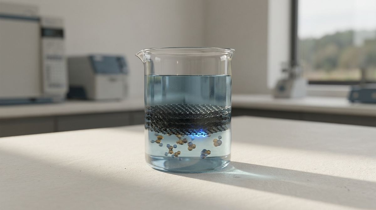
Imagine you are trying to photograph a hummingbird in mid-flight. To capture its wings in perfect detail, you need an incredibly fast shutter speed, or you will end up with a blurry mess. In the world of structural biology, scientists face a similar nightmare when trying to photograph the tiny, moving parts of a cell. They use a technique called Cryo-Electron Microscopy (Cryo-EM) to freeze these biological machines in a thin layer of ice and then blast them with electrons to create a 3D image. However, a major problem arises during the preparation of these samples: the proteins often get caught at the edge of the liquid, causing them to unfold or stick in a single, unhelpful direction. This makes it nearly impossible to see the full structure. A recent breakthrough has provided a way to fix this using a "safety net" made of graphene oxide.
The fundamental challenge in Cryo-EM is the air-water interface, or AWI. When a scientist prepares a sample, they create an incredibly thin film of liquid on a tiny mesh grid. As this liquid is blotted to reach the necessary thickness, a boundary is created between the water and the air. For many proteins, this boundary is a death trap. The surface tension at the air-water interface can physically pull a protein apart, causing it to denature or unfold. Once a protein loses its natural shape, it loses its biological function and becomes useless for scientific study.
Furthermore, even if the protein survives the interface, it often suffers from preferential orientation. This is a phenomenon where proteins, rather than floating randomly in the ice, all land on the surface in the exact same way—for example, always facing "down." To build a high-resolution 3D model, scientists need to see the protein from every possible angle, much like how you need to see a person from the front, side, and back to truly understand their shape. If all the molecules are oriented the same way, there are "blind spots" in the data that prevent high-resolution reconstruction.
Finally, there is the issue of beam-induced motion. When the high-energy electron beam hits the frozen sample, it can cause the ice to move or buckle slightly. Even a tiny amount of movement during the exposure can blur the image, making it impossible to reach the atomic-level resolution required to see individual atoms. The research conducted by Hok Sau Kwong, Alessandro Grinzato, Gian Luca Freiherr von Scholley, Marc‐André Hograindleur, and Eaazhisai Kandiah focuses on solving these specific issues by introducing a supporting layer that manages how the proteins interact with their environment.
The researchers proposed using graphene oxide (GO) as a specialized landing pad for biological molecules. Instead of letting the proteins float freely in a thin layer of water that touches the air, the proteins are allowed to stick to a very thin, transparent sheet of graphene oxide. This sheet acts as a physical barrier that protects the proteins from the destructive forces of the air-water interface. Because the graphene oxide is chemically active, it can hold the proteins in place without the need for extreme chemical modifications that might change the protein's behavior.
To make this work, the researchers had to solve a secondary problem: how to get the graphene oxide to stay on the metal grid during the preparation process. They used a specialized "glue" called polyethyleneimine (PEI). This polymer acts as a bridge, helping the graphene oxide adhere firmly to the grid so it does not get washed away when the liquid is blotted. By using a standard piece of laboratory equipment called the Vitrobot Mark IV, the researchers have demonstrated that you do not need a specialized, multi-million dollar setup to use this advanced technology. You just need a well-optimized protocol.
To understand why this system works, we have to look at the chemistry of graphene oxide. Unlike pure graphene, which is made entirely of carbon atoms in a perfect hexagonal lattice, graphene oxide contains various oxygen-containing functional groups, such as hydroxyl, epoxy, and carboxyl groups. These groups make the surface "hydrophilic," meaning it has a high affinity for water. This is crucial because biological samples are aqueous, and the surface must be able to interact with the water-based protein solution without repelling it.
The introduction of polyethyleneimine (PEI) is the technical masterstroke of this method. PEI is a polymer that carries a high positive charge due to its many amine groups. Graphene oxide, on the other hand, typically carries a negative charge because of its carboxyl groups. When PEI is added to the mix, it creates a strong electrostatic attraction between the graphene oxide and the grid surface. This creates a stable, thin, and uniform layer of graphene oxide that is robust enough to withstand the mechanical stress applied by the Vitrobot's blotting pads during sample preparation.
The Vitrobot Mark IV plays a critical role in maintaining consistency. The device allows for precise control over the humidity, the blotting time, and the force applied to the grid. By maintaining high humidity during the process, the device prevents the liquid from evaporating too quickly, which ensures that the concentration of salts and proteins remains stable. This control is vital for achieving a uniform distribution of the graphene oxide. If the layer is too thick, the electrons cannot pass through it; if it is too thin or patchy, the proteins will fall into the gaps and hit the air-water interface.
The researchers found that this modified drop-cast protocol is highly effective at creating a reliable support layer. Their tests showed that the graphene oxide achieved an average coverage of 55% across the grids. Most importantly, 40% of this coverage consisted of either a single layer (monolayer) or two layers (bilayer) of graphene oxide. In the world of cryo-EM, a monolayer is the ideal scenario because it provides the thinnest possible barrier, allowing electrons to pass through with minimal interference while still providing the necessary support.
The success of the method was proven through the reconstruction of two well-known biological targets: beta-galactosidase and apoferritin. Using this graphene oxide support, the researchers achieved a resolution of 2.3 Å for beta-galactosidase and an incredible 1.9 Å for apoferritin. To put these numbers in perspective, a resolution of 1.9 Å is close to the atomic scale, meaning scientists can see the individual atoms that make up the protein. This level of detail is only possible when the sample is perfectly stabilized and free from the distortions caused by the air-water interface and beam-induced motion.
This result is significant because it democratizes high-resolution structural biology. Previously, achieving these levels of resolution often required highly specialized grids or extremely difficult-to-master sample preparation techniques that were not reproducible across different laboratories. By showing that a standard Vitrobot Mark IV can be used to prepare these advanced grids, the researchers have provided a roadmap that many laboratories can follow.
The ability to achieve atomic resolution means that scientists can see exactly how a drug molecule binds to a protein, or how a mutation in a genetic sequence changes the shape of a vital enzyme. This is the foundation of modern drug discovery. If we can see the structure of a virus or a protein involved in Alzheimer's disease at a 1.9 Å resolution, we can design much more effective medicines. The graphene oxide layer provides the stability needed to capture these high-resolution snapshots without the proteins being ruined by the environment.
While this research is a major step forward, it is not a perfect solution for every problem. One obvious limitation is the coverage percentage. The researchers achieved 55% coverage, which is excellent for many applications, but it means that nearly half of the grid is still bare. In these bare areas, proteins will still encounter the air-water interface, which could lead to inconsistent data if the protein concentration is not perfectly optimized.
Furthermore, while the method works for standard proteins like apoferritin, it remains to be seen how it handles more complex, larger, or more delicate molecular machines. Different proteins have different surface charges and different tendencies to adhere to surfaces. Future research will need to determine if the PEI-enhanced graphene oxide method needs further tuning for specific types of biological samples, such as membrane proteins or large multi-protein complexes, which are notoriously difficult to study.
The implications for real-world science are vast. In the pharmaceutical industry, this method could significantly accelerate the timeline for "structure-based drug design." By providing a more reliable way to see proteins in their native states, pharmaceutical companies can reduce the trial-and-error process involved in developing new medications.
In fundamental biological research, this method could allow scientists to study proteins that were previously considered "un-imageable." Many proteins are difficult to study because they are highly sensitive to their environment or have highly asymmetrical shapes that lead to preferential orientation. The graphene oxide support provides a more diverse chemical landscape, which may allow these challenging proteins to be imaged in multiple orientations, finally revealing their true structures.
If you remember only one thing from this research, let it be this: graphene oxide provides a stable, chemically active "landing pad" that protects delicate biological molecules from the destructive forces of the air-water interface, enabling scientists to see the atomic details of life using standard laboratory equipment.
What is the main purpose of using graphene oxide in cryo-EM? The primary purpose is to provide a stable support for biological molecules, which helps prevent them from sticking to the air-water interface and being damaged or oriented incorrectly. By giving the proteins a surface to adhere to, we can keep them away from the edge of the water droplet.
Why is the air-water interface a problem for scientists? The air-water interface is problematic because the surface tension at the boundary between air and water can physically pull a protein apart, causing it to unfold. Additionally, it often causes proteins to stick in one specific orientation, which prevents scientists from seeing the full 3D structure.
Why do researchers use PEI when preparing these grids? PEI, or polyethyleneimine, acts as a molecular bridge. Because graphene oxide and the metal grid have different electrical charges, the PEI uses electrostatic attraction to glue the graphene oxide onto the grid, ensuring it stays in place during the cleaning and blotting process.
Can this method be used in any laboratory? One of the biggest advantages of this specific research is that it uses the Vitrobot Mark IV, which is a standard piece of equipment in many cryo-EM facilities. This means that laboratories do not need to buy specialized, expensive new machinery to benefit from graphene oxide support.
What does a resolution of 1.9 Å actually mean? In structural biology, resolution refers to the level of detail visible in an image. A resolution of 1.9 Å is extremely high, meaning the image is so clear that researchers can begin to identify the positions of individual atoms within a protein, allowing for very precise scientific conclusions.
The development of a reliable and reproducible method for preparing graphene oxide grids marks a significant milestone in the field of cryo-electron microscopy. By combining the unique chemical properties of graphene oxide with the precision of the Vitrobot Mark IV and the adhesive power of polyethyleneimine, researchers have created a way to bypass the most common obstacles in sample preparation. This method not only protects proteins from the damaging air-water interface but also helps ensure they are oriented correctly for high-resolution imaging. As we continue to refine these techniques, the ability to visualize the machinery of life at an atomic scale will become more accessible, driving breakthroughs in medicine, biology, and beyond.
Serious about B2B integration? Test our premium Pulsed Electrical Resistive Carbon Heating turbostratic graphene in your lab. 100g sample packs available now.
Explore More
Exploring how quantum information geometry provides a new way to understand the complex electrical transport in multicomponent superconductors.

A technical guide for prototyping a non-Faradaic graphene biosensor designed for real-time, label-free quantification of Human Serum Albumin (HSA) using electric double-layer modulation.

Imagine a world where your smartwatch never needs a charger because it stays powered by the warmth of your skin, or where tiny sensors embedded in industrial engines harvest energy from the heat they