Breakthrough Graphene-Collagen Cryogel and Stem Cell Vesicles Offer New Hope for Treating Hypertrophic Scars
Imagine a wound that refuses to heal properly, instead swelling into a raised, thick, and often painful ridge of tissue. These are hypertrophic scars, a common complication of skin injuries, surgeries, or burns that can impact both physical comfort and personal confidence. For decades, medical science has struggled to find a solution that does more than just flatten the surface; we need a way to tell the skin cells to stop behaving aggressively. Recent breakthroughs in material science and regenerative medicine are finally providing a way to communicate directly with these cells. In a significant advancement for regenerative medicine, researchers Mengyuan Jiang, Xiyuan Mao, and L Zhang have developed a dual-action therapeutic platform that combines the structural intelligence of graphene-based cryogels with the biological signaling of stem cell vesicles to fundamentally reshape how the body manages scar formation.
The Problem This Research Is Solving
When the skin is injured, the body initiates a rapid repair mechanism. In a perfect world, this process is controlled and efficient. However, in the case of hypertrophic scars, the repair mechanism goes into overdrive. This overactive response is driven primarily by a specific group of cells known as fibroblasts. In a healthy healing process, fibroblasts produce collagen to bridge the gap in the skin. In hypertrophic scarring, these fibroblasts become hyperactive, transforming into myofibroblasts. These specialized cells are characterized by the expression of alpha-smooth muscle actin, a protein that allows them to contract and pull on the surrounding tissue, contributing to the raised, thickened texture of the scar.
The pathology of hypertrophic scars is marked by a massive overproduction of extracellular matrix components, particularly Type I collagen. This excess collagen builds up in dense, disorganized bundles rather than the neat, organized lattice found in healthy skin. Furthermore, these cells do not stay put; they migrate laterally and proliferate rapidly, creating a self-sustaining cycle of tissue expansion. Current treatments, such as silicone sheets or corticosteroid injections, often focus on managing the symptoms rather than addressing the underlying cellular dysfunction. There is a pressing clinical need for a delivery system that can penetrate the dense scar tissue and deliver active biological agents directly to the problematic fibroblasts to reset their behavior.
The Key Idea in Plain English
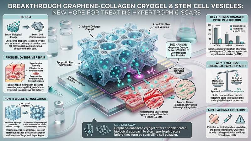
The researchers have proposed a two-part solution that works like a smart, biological sponge. The first part is a specialized material called a graphene-collagen cryogel. Think of this as a highly organized, microscopic sponge made of natural collagen and high-tech graphene. This sponge is not just a filler; it is engineered to have large, interconnected tunnels that allow it to absorb and hold onto "messenger" particles.
The second part of the solution is a collection of tiny biological packages called apoptotic vesicles, specifically derived from adipose-derived stem cells. These vesicles are released when stem cells undergo a natural, controlled process of cell death. These vesicles are essentially tiny cargo ships carrying instructions that tell the skin cells to calm down. By loading these "instruction ships" into the graphene-collagen sponge, the researchers created a system that can stay at the site of a scar and slowly release these calming signals, teaching the hyperactive scar cells to return to a normal, non-scarring state.
How the Graphene-Based System Works
To understand why this system is so effective, we must look at the chemistry of the cryogel and the unique properties of graphene. Traditional hydrogels, which are often used in medicine, typically have very small, microscopic pores. These small pores make it difficult for large biological molecules or vesicles to move through the material and even harder to load them effectively. The researchers used a process called cryogelation to solve this. By freezing the collagen solution during the formation process, they created ice crystals that acted as physical templates. As the ice crystals grew, they pushed the collagen molecules aside, creating large, interconnected pores. Once the ice melts, these empty spaces remain, leaving behind a highly porous, three-dimensional scaffold.
This high porosity is critical for two reasons. First, it allows the gel to be easily loaded with a high concentration of apoptotic vesicles. Second, it provides a physical highway that allows the vesicles to be released steadily over time. This is where graphene becomes indispensable. Graphene is a single layer of carbon atoms arranged in a hexagonal lattice, providing an incredibly high surface-area-to-volume ratio. When graphene is incorporated into the collagen matrix, it creates a vast network of surfaces that have a high affinity for the apoptotic vesicles.
The interaction between the graphene and the vesicles is a matter of selective adsorption. Because of the unique chemical interface provided by the graphene flakes, the vesicles are not just floating loosely in the gel; they are strategically adsorbed onto the graphene surfaces. This ensures that the vesicles are not washed away by bodily fluids immediately after application. Instead, the graphene acts as a reservoir, controlling the release of the vesicles through a steady diffusion process. This controlled release is vital because it maintains a consistent therapeutic concentration of the biological signals around the scar tissue, ensuring the fibroblasts receive continuous instructions to stop producing excess collagen.
What the Researchers Found
The study yielded highly significant results across several biological markers. When the researchers tested the graphene-collagen cryogel (G-GEL(C)) alone on hypertrophic scar fibroblasts, they observed a dramatic reduction in the proteins responsible for scar formation. Specifically, there was a significant downregulation of COL1A1, the primary type of collagen found in scars, as well as alpha-SMA, the marker for the aggressive myofibroblasts. The study also noted a significant reduction in Vimentin, a protein involved in cell structure and movement. By reducing these proteins, the G-GEL(C) effectively stripped the fibroblasts of their ability to build thick tissue and contract the skin.
The presence of the ASCs-ApoVs (the stem cell vesicles) amplified these effects. When the vesicles were added to the mix, they further suppressed the expression of COL1A1 and alpha-SMA at both the protein and mRNA levels. This suggests that the vesicles are successfully interfering with the genetic instructions of the cells, telling them to stop producing the building blocks of the scar. The combination of the gel's physical presence and the vesicles' chemical signaling led to a significant decrease in cell proliferation and lateral migration, meaning the cells stopped multiplying and stopped spreading across the wound.
In vivo testing, which involves living organisms, confirmed these findings. When the G-GEL(C) loaded with vesicles was applied, it significantly reduced the Scar Elevation Index (SEI), which is a clinical measurement of how much a scar protrudes above the skin surface. Furthermore, the research showed an increase in M2 macrophages at the site of the scar. In the context of wound healing, macrophages are the "cleanup crew" of the immune system. While M1 macrophages promote inflammation, M2 macrophages are responsible for tissue remodeling and anti-inflammatory responses. By shifting the environment toward an M2 macrophage presence, the treatment helps transition the wound from a state of aggressive inflammation to a state of organized, calm remodeling.
Why the Result Matters
This research is important because it shifts the paradigm of scar treatment from mechanical suppression to biological regulation. For a long time, the medical community has treated hypertrophic scars as a problem of "too much of a good thing"—too much collagen, too much cell activity. Most existing treatments simply try to flatten the result. This new approach targets the "why" behind the scar. By using a graphene-based delivery system, we can deliver complex biological instructions with a precision that was previously impossible.
Furthermore, the use of apoptotic vesicles is a brilliant way to leverage the body's own signaling pathways. Using whole stem cells in therapy can be risky, as they can be difficult to control once they are injected. However, using the vesicles they release is much safer, as the vesicles contain the beneficial signals without the risks associated with living cell transplantation. This makes the therapy more stable, easier to manufacture, and much more predictable in a clinical setting. The ability to combine a structural material (the cryogel) with a biological signal (the vesicles) represents a new frontier in combinatorial therapy.
Limitations and What Still Needs Testing
While these results are highly promising, it is important to approach them with scientific caution. It is critical to understand that this research has been conducted primarily in laboratory settings and in animal models. While animal models are a vital step in the scientific process, they do not always perfectly replicate the complexities of human skin or the human immune system. The way a rodent heals a scar is fundamentally different from the way a human heals a scar.
Therefore, the transition from a successful laboratory experiment to a safe and effective human treatment requires extensive clinical trials. Researchers must still determine the optimal dosage of vesicles, the ideal frequency of gel application, and the long-term safety of graphene-collagen scaffolds within the human body. There is also the question of how the body will eventually clear the graphene-collagen scaffold once its job is done. While the cryogel is designed to be biocompatible, the long-term metabolic fate of these synthetic-natural hybrid materials must be thoroughly investigated before this can become a standard medical procedure.
Real-World Applications
The potential applications for this graphene-collagen-vesicle platform extend far beyond just aesthetic scar reduction. In the field of regenerative medicine, this technology could be adapted for various types of wound management. For patients with chronic wounds, such as diabetic ulcers, which often fail to heal due to a persistent inflammatory state, similar "smart" scaffolds could be used to deliver anti-inflammatory signals and promote healthy tissue growth.
Additionally, this technology could be used in reconstructive surgery to minimize the visibility of scars from complex procedures, such as breast reconstruction or neurological surgeries. Because the system can be tailored with different types of vesicles or different concentrations of graphene, it offers a customizable approach to wound healing. As our ability to engineer graphene and biological vesicles improves, we may see this platform used to treat a wide variety of fibrotic diseases, where the body's healing process has gone dangerously off the rails.
If You Remember One Thing
If you take away only one piece of information from this research, let it be this: the future of scar treatment lies in "smart" materials that don't just cover a wound, but actively communicate with the cells to prevent the biological mistakes that lead to hypertrophic scarring.
FAQ
What exactly is a hypertrophic scar?
A hypertrophic scar is a raised, thick, and often red scar that forms when the body's healing process goes into overdrive. Unlike a normal scar, which settles down once the wound is closed, a hypertrophic scar continues to grow and produce excess collagen, resulting in a bump that stays within the boundaries of the original injury.
Why is graphene used in this research?
Graphene is used because of its incredible surface area and its unique ability to interact with other molecules. In this study, graphene acts as a high-tech storage unit, allowing the medical gel to hold onto therapeutic vesicles and release them slowly and steadily, rather than all at once.
What are apoptotic vesicles?
When stem cells undergo a natural, programmed cell death called apoptosis, they release tiny bubbles or vesicles. These vesicles carry various chemical signals that can influence the behavior of neighboring cells. In this research, those signals are used to tell problematic scar cells to stop being so aggressive.
What is the difference between a hydrogel and a cryogel?
A hydrogel is a jelly-like substance that is very good at holding water, but it usually has very tiny pores. A cryogel is made using a freezing process that creates much larger, interconnected tunnels within the material. These larger tunnels make it much easier to load the gel with medicinal particles and allow them to move through the gel more effectively.
Does this mean scars will never be a problem again?
Not exactly. While this research is a massive leap forward, it is still in the early stages of testing. This technology represents a new way to treat scars, but it must undergo many more years of testing in humans to ensure it is safe, effective, and consistent for everyone.
Conclusion
The convergence of nanotechnology and cell biology is opening doors that were previously locked. By integrating the structural advantages of graphene-based cryogels with the biological intelligence of stem cell vesicles, researchers have created a potent tool for modulating the complex processes that lead to hypertrophic scars. This combinatorial approach—addressing both the physical structure of the scar and the cellular behavior driving it—marks a significant step toward a more sophisticated, biological approach to wound healing. As we move from the laboratory to clinical trials, the ability to "reprogram" how our skin heals could change the lives of millions of people.
Evaluate Our Quality
Serious about B2B integration? Test our premium Pulsed Electrical Resistive Carbon Heating turbostratic graphene in your lab. 100g sample packs available now.