Graphene-Powered Terahertz Sensors for Early Breast Cancer Detection
Imagine a world where breast cancer is detected not through uncomfortable compression or invasive biopsies, but by a portable sensor that can see the earliest signs of malignancy without ever breaking the skin. For millions of women, early detection is the difference between a manageable condition and a life-threatening battle. However, current screening methods often struggle to identify the smallest tumors or accurately stage the disease without invasive procedures. This is where the intersection of nanotechnology and electromagnetic physics offers a breakthrough. Recent research led by Mahesh Kumar Aghwariya, Vanshika Goel, Sasmita Dash, Sudhi Agarwal Kamboj, and Tushar Goel has introduced a miniaturized biosensor that leverages the unique properties of graphene to detect breast cancer at its earliest stages and help determine how far it has spread.
The Problem This Research Is Solving
Breast cancer is one of the leading causes of mortality among women globally, and its lethality is closely tied to how late it is discovered. The gold standard for screening has long been mammography, but this method has significant drawbacks. It involves ionizing radiation, which can be harmful over repeated exposures, and it often requires uncomfortable tissue compression. Furthermore, mammography can sometimes miss very small tumors or struggle to differentiate between benign cysts and malignant growths in dense breast tissue.
Beyond simple detection, doctors need to determine the TNM stage of the cancer. The T stands for the size and extent of the primary tumor, N refers to whether the cancer has spread to nearby lymph nodes, and M indicates if the cancer has metastasized to distant organs. Determining these factors usually requires invasive biopsies or complex imaging that can be expensive and time-consuming. There is a critical need for a noninvasive, portable device that can provide high-resolution information about tumor size and nodal involvement in real-time, allowing for faster and more accurate treatment planning.
The Key Idea in Plain English
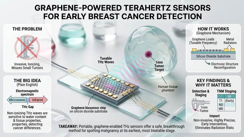
The researchers proposed using terahertz (THz) waves, which are a form of electromagnetic radiation that sits between microwaves and infrared light. THz waves are particularly useful for medical imaging because they are non-ionizing, meaning they do not damage DNA like X-rays do. Most importantly, THz waves are incredibly sensitive to water content and the dielectric properties of biological tissues. Since cancerous tumors typically have higher water concentrations and different cellular structures than healthy breast tissue, they reflect THz waves differently.
To make this detection highly precise and portable, the team integrated graphene into the sensor design. Graphene is a single layer of carbon atoms arranged in a hexagonal lattice, and it possesses an extraordinary ability to change its electrical conductivity based on an applied voltage. By using graphene as a tunable component, the researchers created a sensor that can be adjusted to different frequencies. This adjustability allows the device to scan through various depths and tissue types, effectively acting like a tunable radio that can hone in on the specific signature of a cancerous tumor or a compromised lymph node.
How the Graphene-Based System Works
The biosensor is constructed using a silicon dioxide substrate, which provides a stable, insulating base. On top of this substrate, the researchers placed metal radiators and graphene loads. The interaction between these components creates a resonant system capable of emitting and receiving THz waves. The most critical aspect of this design is the use of graphene to achieve frequency reconfiguration.
In traditional sensors, the operating frequency is fixed by the physical dimensions of the device. However, graphene possesses a unique electronic structure characterized by a linear dispersion relation near the Dirac point. This means that its conductivity is not fixed; instead, it can be modulated by changing the chemical potential of the electrons within the graphene layer. By applying an external DC bias voltage, the researchers can shift the Fermi level of the graphene. This change in chemical potential directly alters the surface conductivity of the graphene, which in turn shifts the resonant frequency of the entire sensor. This allows the device to operate across a wide range, specifically from 3.13 to 5.72 THz.
This tunability is essential because different types of tissue and tumors respond differently to various frequencies. By sweeping through this range, the sensor can optimize its sensitivity to detect tumors of varying sizes. The device also achieves high directivity and gain, meaning it can focus its electromagnetic energy into a narrow beam. This focusing effect is caused by the precise geometry of the metal radiators and the high carrier mobility of graphene, which ensures that signal loss is minimized as the wave travels toward the target tissue.
What the Researchers Found
To test their device, the team developed a sophisticated human breast phantom model. This phantom was designed to mimic the exact dielectric properties of real human breast tissue and nearby lymph nodes in the THz regime. By simulating how waves bounce off different densities of matter, they could evaluate the sensor's accuracy without risking human subjects.
The results were impressive. The biosensor was able to detect tumors ranging from as small as 1 millimeter up to 10 millimeters. This ability to spot a 1mm tumor is significant, as it represents the very early stages of cancer development. The researchers also found that the sensor's performance remained robust even when the angle or position of the device relative to the tissue changed, which is vital for real-world clinical use where a patient may move slightly during a scan.
Furthermore, the sensor proved capable of detecting cancerous cells that had migrated to nearby lymph nodes. By analyzing the reflectometry data—essentially measuring how much of the THz wave was reflected back to the sensor after hitting the tissue—the team could distinguish between healthy lymph nodes and those containing malignant cells. This means the device can potentially automate much of the TNM staging process, providing clinicians with a noninvasive map of tumor size and nodal involvement.
Why the Result Matters
The implications of this research are profound because it addresses both safety and precision. Because THz waves are non-ionizing, this technology eliminates the radiation risks associated with X-ray based mammography. This makes it a safer option for frequent screening, which is often necessary for women with a high genetic predisposition to breast cancer.
Moreover, the miniaturization of the sensor opens the door to portable diagnostic tools. Instead of requiring a massive, expensive imaging suite in a hospital, a graphene-based THz sensor could eventually be integrated into handheld devices. This would democratize access to early cancer screening, allowing clinics in rural or underserved areas to perform high-quality diagnostics. The ability to detect tumors at the 1mm scale significantly increases the likelihood of successful treatment, as early-stage cancers are far more responsive to therapy and less likely to have metastasized.
Limitations and What Still Needs Testing
While the results are promising, it is important to note that this research was conducted using a phantom model. A breast phantom, while scientifically accurate in terms of dielectric constants, cannot fully replicate the complex biological variability of a living human being. Factors such as blood flow, varying skin thickness, and different hormonal states can all influence how THz waves interact with tissue. Therefore, the next critical step is to move from phantom models to controlled in vivo human trials to validate these findings.
Another limitation is the penetration depth of THz radiation. THz waves are highly absorbed by water, which means they cannot penetrate very deep into the body. While this is excellent for detecting tumors near the surface or in a biopsy sample, it may limit the sensor's ability to find very deep-seated tumors without supplementary imaging. Further research is needed to optimize the frequency range and power output to maximize penetration while maintaining safety and resolution.
Real-World Applications
If successfully transitioned to clinical settings, this technology could serve several roles. First, it could act as a primary screening tool for women with dense breast tissue, where traditional mammograms often fail. Second, it could be used during surgical procedures to provide real-time guidance. Surgeons could use a handheld version of the sensor to ensure they have removed all cancerous tissue while sparing as much healthy tissue as possible, a process known as checking the surgical margins.
Additionally, the sensor could be used to monitor the effectiveness of chemotherapy. Since chemo often changes the water content and cellular structure of a tumor as it shrinks, the THz biosensor could provide a noninvasive way to track whether a tumor is responding to treatment in real-time, allowing doctors to adjust dosages or switch medications more quickly.
If You Remember One Thing
The most important takeaway is that by combining the tunable electrical properties of graphene with non-ionizing terahertz waves, researchers have created a sensor capable of detecting breast cancer tumors as small as 1mm and staging the disease noninvasively, potentially replacing more painful and risky screening methods.
FAQ
What is terahertz radiation and is it safe?
Terahertz radiation occupies the space on the electromagnetic spectrum between microwaves and infrared light. Unlike X-rays, terahertz waves are non-ionizing, which means they do not have enough energy to strip electrons from atoms or damage DNA. This makes them exceptionally safe for medical imaging of soft tissues.
How does graphene make the sensor better?
Graphene acts as a tunable conductor. By applying a small amount of voltage, the researchers can change how electrons move within the graphene layer, which shifts the frequency at which the sensor operates. This allows the device to be adjusted to find the best signal for different tumor sizes or tissue depths.
What does TNM staging mean in this context?
TNM is a system used to describe the extent of cancer: T for Tumor size, N for Node involvement (whether it reached lymph nodes), and M for Metastasis (whether it spread to other organs). This sensor helps determine the T and N stages by identifying tumor size and nodal health noninvasively.
Can this sensor replace mammograms entirely?
While it offers many advantages, such as being radiation-free and highly sensitive to small tumors, it is more likely to complement existing tools. Because terahertz waves have limited penetration depth, they may be used alongside other imaging techniques to provide a more complete picture of a patient's health.
Will this device be available in hospitals soon?
The research is currently at the stage of successful phantom model testing. Before it becomes a commercial medical device, it must undergo rigorous human clinical trials to ensure accuracy across diverse patient populations and receive regulatory approval.
Conclusion
The work by Aghwariya and colleagues represents a significant leap forward in the application of 2D materials for healthcare. By harnessing the unique electronic properties of graphene, they have developed a tool that is not only sensitive and precise but also adaptable. The ability to tune the sensor's frequency allows for a level of diagnostic flexibility that was previously unattainable with fixed-frequency devices. As this technology moves from the laboratory and phantom models into clinical trials, it holds the potential to transform breast cancer screening from a stressful, invasive experience into a simple, safe, and highly accurate routine. In the fight against cancer, time is the most valuable resource, and tools that can detect malignancy at the 1mm scale provide patients with the best possible chance for a full recovery.
Evaluate Our Quality
Serious about B2B integration? Test our premium Pulsed Electrical Resistive Carbon Heating turbostratic graphene in your lab. 100g sample packs available now.