
Probing 2D Metals: Unlocking Electronic Secrets with Raman Spectroscopy
Researchers reveal how breaking inversion symmetry in 2D metals enables electronic Raman scattering, a powerful, non-destructive tool for characterizing...
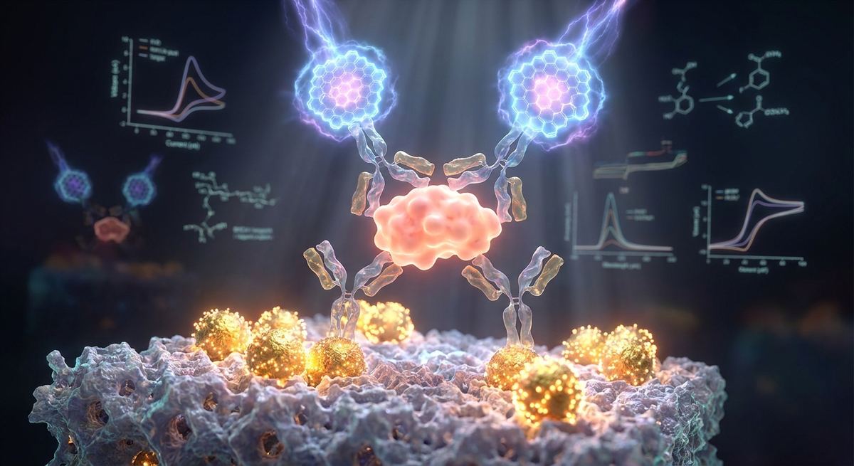
Research conducted by: Lin Wang, Yanyan Wang, Chunchun Chen, Y Li, Huiming Dong, Tingjing Yao, Gongsheng Jin, Zhenjie Wang
The groundbreaking research conducted by this esteemed team of scientists represents a monumental leap forward in the field of clinical diagnostics and nanomaterial engineering. Their comprehensive study meticulously details the development of a highly sensitive sandwich-type electrochemical immunosensor designed specifically for the quantitative detection of breast cancer susceptibility protein 1. By ingeniously combining zeolitic imidazolate framework-8 decorated with gold nanoparticles and nitrogen-doped graphene quantum dots, the research team has successfully engineered a platform that overcomes the traditional limitations of protein biomarker detection. Their work not only provides a highly reliable analytical tool for the early screening of breast cancer but also establishes a versatile methodological framework that can be adapted for a wide array of disease-related proteins. The dedication to optimizing the sensing interface, rigorously validating the analytical performance, and ensuring the sensor's viability in complex human serum highlights the exceptional caliber of this scientific endeavor.
Breast cancer remains one of the most prevalent and life-threatening malignancies affecting women globally. The early and accurate detection of this disease is paramount for improving patient survival rates and guiding effective therapeutic interventions. Among the various biomarkers associated with breast cancer, breast cancer susceptibility protein 1, universally known as BRCA1, plays a critical role. BRCA1 is a tumor suppressor protein that is fundamentally involved in the repair of double-strand DNA breaks through a process known as homologous recombination. When the BRCA1 gene is mutated, or when the expression levels of the BRCA1 protein are abnormally altered, the cellular DNA repair mechanisms are severely compromised. This genomic instability significantly elevates the risk of developing breast and ovarian cancers.
Traditionally, the assessment of BRCA1 has heavily relied on genomic sequencing to identify mutations at the DNA level. While genetic testing is invaluable for determining hereditary predisposition, the real-time monitoring of BRCA1 protein levels in physiological fluids offers a complementary and highly dynamic approach to cancer screening and prognosis. The concentration of the BRCA1 protein in blood or serum can serve as a direct indicator of physiological anomalies. However, detecting this protein at the extremely low concentrations typical of early-stage pathogenesis presents a formidable analytical challenge.
Conventional protein detection methodologies, such as enzyme-linked immunosorbent assays and mass spectrometry, have long been the gold standard in clinical laboratories. Despite their reliability, these techniques are often hindered by inherent limitations. They typically require sophisticated, expensive instrumentation, highly trained personnel, and extensive sample preparation times. Furthermore, their sensitivity thresholds are sometimes inadequate for detecting the minute quantities of biomarkers present in the nascent stages of disease. Consequently, there is an urgent and clinical demand for the development of alternative diagnostic platforms that are not only rapid, cost-effective, and user-friendly but also capable of delivering ultrasensitive and highly specific detection of BRCA1.
Electrochemical immunosensors have emerged as a highly promising solution to this diagnostic bottleneck. By translating the highly specific biological recognition event between an antibody and its target antigen into a measurable electrical signal, these sensors offer a unique combination of high sensitivity, rapid response, and potential for miniaturization. The research team recognized this potential and embarked on a mission to push the boundaries of electrochemical sensing by leveraging the synergistic properties of advanced nanomaterials.
The foundation of this innovative immunosensor lies in the strategic utilization of zeolitic imidazolate framework-8, a prominent member of the metal-organic framework family. Metal-organic frameworks are highly ordered crystalline porous materials constructed from metal ions or metal clusters coordinated to organic ligands. ZIF-8, specifically, is synthesized through the coordination of zinc ions with 2-methylimidazole linkers. This unique structural composition endows ZIF-8 with exceptional physicochemical properties, including an exceptionally high specific surface area, tunable pore sizes, and remarkable chemical and thermal stability.
In the context of biosensor design, the high specific surface area of ZIF-8 is of paramount importance. It provides a vast, three-dimensional scaffold capable of accommodating a massive number of bio-recognition elements, such as capture antibodies. The dense immobilization of these capture antibodies is directly correlated with the sensor's ability to capture the target analyte, thereby fundamentally dictating the ultimate sensitivity of the device. However, despite its impressive structural attributes, pristine ZIF-8 possesses a critical flaw when applied to electrochemical sensing: it exhibits notoriously poor electrical conductivity. This insulating nature impedes the efficient transfer of electrons between the biological recognition event and the underlying electrode surface, severely dampening the electrochemical signal.
To circumvent this limitation, the researchers executed a brilliant modification strategy by decorating the ZIF-8 matrix with gold nanoparticles. The integration of gold nanoparticles into the ZIF-8 framework, creating the AuNPs@ZIF-8 nanocomposite, serves multiple synergistic purposes. First and foremost, gold nanoparticles are excellent electrical conductors. Their presence within the porous network of ZIF-8 creates highly efficient electron conduction pathways, dramatically accelerating the rate of electron transfer across the sensing interface. This enhanced conductivity is crucial for generating a strong, easily measurable electrochemical response.
Furthermore, gold nanoparticles exhibit exceptional biocompatibility and possess a high affinity for biomolecules containing amine and thiol functional groups. This characteristic facilitates the robust and stable immobilization of the BRCA1 capture antibodies onto the nanocomposite surface via strong chemical bonding. The resulting AuNPs@ZIF-8 composite not only retains the massive surface area required for high-density antibody loading but also transforms the previously insulating framework into a highly conductive, bio-friendly microenvironment optimized for electrochemical transduction.
While the AuNPs@ZIF-8 nanocomposite provides an optimized platform for capturing the BRCA1 target, achieving ultrasensitive detection requires a robust signal amplification strategy. In a sandwich-type immunosensor, this is typically accomplished using signal labels attached to a secondary antibody. For this critical role, the researchers turned to nitrogen-doped graphene quantum dots, a cutting-edge class of zero-dimensional carbon nanomaterials.
Graphene quantum dots are essentially nanometer-sized fragments of graphene that exhibit quantum confinement and edge effects. They retain the exceptional electrical conductivity and large surface area of bulk graphene while introducing new, tunable electronic properties due to their incredibly small size. To further enhance their performance as electrochemical signal labels, the researchers introduced nitrogen atoms into the carbon lattice of the quantum dots, a process known as nitrogen doping.
The incorporation of nitrogen atoms fundamentally alters the electronic structure of the graphene quantum dots. Because nitrogen is more electronegative than carbon, its presence induces a redistribution of electron density within the carbon lattice. This creates localized areas of high positive charge density, which serve as highly active catalytic sites. The nitrogen doping also increases the density of states near the Fermi level, facilitating faster interfacial electron transfer kinetics. Consequently, nitrogen-doped graphene quantum dots exhibit significantly superior electrocatalytic activity compared to their undoped counterparts.
In the design of the immunosensor, these highly active nitrogen-doped graphene quantum dots are conjugated with secondary antibodies to form the signal nanoprobe. When the secondary antibody binds to the BRCA1 target, it brings a massive payload of highly electroactive quantum dots into close proximity with the electrode surface. Upon the application of a specific electrical potential, these quantum dots facilitate a rapid and massive transfer of electrons, generating a heavily amplified electrochemical signal. This powerful signal amplification mechanism is the key to unlocking the sensor's ability to detect BRCA1 at extraordinarily low concentrations.
The construction of the electrochemical immunosensor is a highly intricate, stepwise process that demands meticulous precision to ensure optimal performance. The researchers utilized a glassy carbon electrode as the foundational base for the sensor. The assembly process begins with the rigorous polishing and cleaning of the glassy carbon electrode to provide a pristine, mirror-like surface, free from any contaminants that could interfere with electron transfer.
Following the preparation of the electrode, a carefully measured suspension of the AuNPs@ZIF-8 nanocomposite is drop-cast onto the electrode surface and allowed to dry. This step establishes the highly conductive, high-surface-area substrate. Next, a solution containing the primary capture antibodies, specifically targeted against BRCA1, is introduced to the modified electrode. The capture antibodies anchor themselves securely to the gold nanoparticles within the composite through strong interactions between the gold surface and the functional groups on the antibody proteins.
Once the capture antibodies are firmly immobilized, it is imperative to block any remaining exposed active sites on the electrode surface. This is achieved by incubating the electrode in a solution of bovine serum albumin. The albumin molecules act as a passivating layer, binding to any unoccupied areas and preventing the non-specific adsorption of other proteins during subsequent steps. This blocking phase is absolutely critical for minimizing background noise and ensuring the sensor's high specificity.
The sensor is now primed for the detection phase. The sample solution suspected of containing the BRCA1 antigen is applied to the sensing interface. If BRCA1 is present, it will be specifically recognized and captured by the immobilized primary antibodies. Finally, the signal nanoprobe, consisting of the secondary antibodies conjugated with the nitrogen-doped graphene quantum dots, is introduced. The secondary antibodies bind to a different epitope on the captured BRCA1 antigen, effectively sandwiching the target protein between the primary and secondary antibodies. This completes the formation of the immunocomplex and positions the highly electroactive quantum dot labels right at the sensing interface, ready for electrochemical interrogation.
To verify the successful, stepwise assembly of the immunosensor and to interrogate its analytical performance, the researchers employed two powerful electrochemical techniques: cyclic voltammetry and electrochemical impedance spectroscopy. These techniques are performed using a standard electrochemical cell containing a reversible redox probe, typically a mixture of potassium ferricyanide and potassium ferrocyanide, dissolved in a supporting electrolyte solution.
Cyclic voltammetry involves sweeping the potential applied to the electrode back and forth between two predetermined limits and measuring the resulting current. This technique provides a clear visual representation of the electron transfer processes occurring at the electrode surface. As the sensor is assembled step by step, the deposition of non-conductive biomolecules, such as antibodies and blocking agents, creates a physical and electrostatic barrier on the electrode surface. This barrier hinders the diffusion of the redox probe to the underlying conductive substrate, resulting in a progressive decrease in the peak currents observed in the cyclic voltammograms. The researchers documented these expected decreases at each stage of fabrication, providing compelling evidence that the biological layers were successfully immobilized.
Electrochemical impedance spectroscopy offers a complementary and even more sensitive method for characterizing the sensing interface. This technique applies a small alternating current signal over a range of frequencies and measures the complex impedance of the system. The resulting data is typically visualized using a Nyquist plot, which consists of a semicircular portion at higher frequencies and a linear portion at lower frequencies. The diameter of the semicircle corresponds directly to the charge transfer resistance of the electrode surface.
Similar to the cyclic voltammetry results, the charge transfer resistance increases sequentially with the addition of each biological layer. The bare glassy carbon electrode exhibits a very small semicircle, indicating rapid electron transfer. The deposition of the highly conductive AuNPs@ZIF-8 nanocomposite often facilitates even faster electron transfer. However, the subsequent immobilization of the capture antibodies, the bovine serum albumin blocking layer, and the bulky BRCA1 antigen progressively insulate the surface, leading to dramatic increases in the diameter of the Nyquist semicircle. The systematic monitoring of these impedance changes confirmed beyond a doubt that the sandwich immunocomplex was successfully constructed on the electrode surface.
With the sensor's architecture verified, the research team proceeded to evaluate its analytical performance for the quantitative detection of BRCA1. For these critical measurements, they utilized differential pulse voltammetry, an advanced electrochemical technique that minimizes the background capacitive charging current, thereby offering significantly higher sensitivity than standard linear sweep methods.
The sensor was incubated with varying concentrations of the BRCA1 antigen, ranging from incredibly dilute solutions to much higher concentrations. The differential pulse voltammetry responses revealed a clear, systematic relationship: as the concentration of BRCA1 increased, the magnitude of the electrochemical signal generated by the nitrogen-doped graphene quantum dot labels also increased.
Crucially, the researchers discovered a highly linear relationship between the change in peak current and the logarithm of the BRCA1 concentration over an astonishingly wide dynamic range, spanning from 1.0 picogram per milliliter to 50 nanograms per milliliter. The calibration curve yielded a linear regression equation of delta I equals 3.58 times the log of concentration plus 7.21, with an exceptional correlation coefficient of 0.9985. This high degree of linearity indicates that the sensor provides highly reliable and predictable quantitative data across a vast spectrum of target concentrations.
Even more impressive is the sensor's limit of detection, which was calculated to be a mere 0.3 picograms per milliliter. This ultrasensitive detection capability represents a significant improvement over many conventional commercial assays and previously reported diagnostic platforms. The ability to detect BRCA1 at such trace levels is a major breakthrough, as it opens the door for the extremely early identification of biomarker fluctuations, potentially allowing for medical intervention long before physical symptoms manifest or tumors become visible on traditional imaging scans.
For an immunosensor to be truly viable for clinical applications, it must not only be highly sensitive but also exceptionally selective, stable, and capable of operating in complex biological matrices. Human blood and serum are intricate mixtures containing thousands of different proteins, metabolites, and other biochemical compounds that can easily interfere with analytical measurements, leading to false-positive or false-negative results.
To rigorously assess the sensor's selectivity, the researchers tested its response to several potential interfering substances commonly found in human serum, including ascorbic acid, uric acid, human serum albumin, and various globulins, at concentrations much higher than that of the target BRCA1. Remarkably, the immunosensor exhibited a negligible response to these interferents, with the variation in signal remaining well below 5 percent. This high degree of specificity is attributed to the inherent precision of the antigen-antibody biological recognition event and the effective blocking strategy employed during sensor fabrication.
The long-term stability and reproducibility of the sensor were also thoroughly investigated. Reproducibility, a measure of the sensor's consistency across multiple fabrications, was evaluated by constructing several independent electrodes under identical conditions. The relative standard deviation of their responses was a highly acceptable 3.8 percent, demonstrating the reliability of the manufacturing protocol. Furthermore, when stored under appropriate refrigerated conditions, the immunosensor retained an impressive 92.5 percent of its initial signal response after 30 days, indicating excellent long-term stability and shelf life, which are crucial factors for commercialization and routine clinical use.
The ultimate test of the sensor's clinical utility was its application in real human serum samples. The researchers performed spike-and-recovery experiments, where known concentrations of BRCA1 were added to healthy human serum, and the sensor was used to measure the total concentration. The calculated recovery rates were outstanding, ranging from 96.8 percent to 104.2 percent. These results definitively prove that the complex biochemical environment of human serum does not significantly impede the sensor's performance. The platform demonstrated remarkable robustness and accuracy, confirming its immense potential as a practical, highly sensitive diagnostic tool for real-world clinical environments.
The development of this AuNPs@ZIF-8 and N-GQD-based electrochemical immunosensor represents a paradigm shift in the approach to clinical diagnostics and personalized oncology. By successfully marrying the structural advantages of metal-organic frameworks with the extraordinary electronic properties of carbon quantum dots, the research team has created a diagnostic platform that vastly outperforms traditional methodologies in terms of sensitivity, speed, and operational simplicity.
The implications of this technology extend far beyond the detection of BRCA1. The modular nature of the sandwich immunosensor design means that by simply substituting the primary and secondary antibodies, this platform can be readily adapted to detect a vast array of other critical disease biomarkers, ranging from cardiac troponins for heart attack diagnosis to specific viral antigens for infectious disease monitoring.
Furthermore, the inherent characteristics of electrochemical sensors make them ideal candidates for integration into miniaturized, point-of-care diagnostic devices. Unlike bulky laboratory equipment, electrochemical platforms can be easily miniaturized and interfaced with microfluidic systems and portable electronics, such as smartphones. This paves the way for the development of highly accessible, rapid diagnostic tools that can be deployed in resource-limited settings, remote clinics, or even for at-home patient monitoring. The research team's pioneering work has not only provided a powerful new weapon in the fight against breast cancer but has also laid down a versatile technological foundation that will undoubtedly drive the next generation of ultrasensitive biomedical diagnostics.
What is a sandwich-type electrochemical immunosensor?
A sandwich-type electrochemical immunosensor is a specialized diagnostic device that uses a dual-antibody system to detect a specific target protein. It involves a capture antibody immobilized on an electrode surface that binds to the target antigen from a sample. A secondary antibody, equipped with a signal-generating label, is then introduced to bind to a different site on the same antigen. This creates a "sandwich" structure. The label on the secondary antibody generates an electrical signal when a voltage is applied, and the strength of this signal is directly proportional to the amount of target protein present in the sample.
Why is BRCA1 an important biomarker?
BRCA1, or breast cancer susceptibility protein 1, is a crucial protein involved in repairing damaged DNA and maintaining the genetic stability of cells. When the BRCA1 gene is mutated, or when the protein levels are abnormal, the body's ability to fix DNA breaks is impaired, significantly increasing the likelihood of developing breast and ovarian cancers. Monitoring the levels of the BRCA1 protein in bodily fluids can serve as an early warning system, helping clinicians identify patients at high risk and monitor the progression or recurrence of the disease.
How do gold nanoparticles improve ZIF-8?
ZIF-8 is a highly porous metal-organic framework with a massive surface area, making it excellent for holding large amounts of capture antibodies. However, ZIF-8 is basically an electrical insulator, which is detrimental for an electrochemical sensor that relies on electron flow. By decorating the ZIF-8 with gold nanoparticles, researchers solve this problem. The gold acts as tiny, highly conductive wires throughout the framework, facilitating rapid electron transfer. Additionally, the gold provides excellent chemical anchor points for attaching the biological antibodies securely to the sensor surface.
What role do nitrogen-doped graphene quantum dots play?
In this sensor, nitrogen-doped graphene quantum dots serve as the powerful signal labels attached to the secondary antibodies. Graphene quantum dots are tiny fragments of carbon that are highly conductive. By doping them with nitrogen atoms, researchers alter their electronic structure, making them highly active electrocatalysts. When the secondary antibody binds to the target, it brings these quantum dots to the electrode. Upon electrical stimulation, the doped quantum dots generate a massive, amplified electrical current, allowing the sensor to detect even trace, picogram-level amounts of the BRCA1 protein.
Can this sensor be used for other diseases?
Yes, the fundamental architecture of this biosensor is highly versatile and adaptable. While this specific study focused on detecting the BRCA1 protein for breast cancer screening, the underlying technology can be repurposed. By simply swapping out the primary capture antibodies and the secondary labeled antibodies for ones that specifically recognize different disease biomarkers, the same AuNPs@ZIF-8 and N-GQD platform can be used to detect markers for prostate cancer, infectious diseases, cardiovascular events, and various other critical health conditions.
The brilliant engineering of this sandwich-type electrochemical immunosensor marks a significant milestone in nanomedicine and clinical diagnostics. By harnessing the high surface area of gold nanoparticle-decorated ZIF-8 and the powerful signal amplification of nitrogen-doped graphene quantum dots, the researchers have achieved an extraordinary detection limit for the BRCA1 biomarker. This platform effectively bridges the gap between advanced nanomaterial science and critical clinical needs, offering a rapid, highly sensitive, and robust alternative to conventional testing methods. As this technology transitions from the laboratory toward commercial point-of-care applications, it holds the profound potential to revolutionize early cancer screening, ultimately facilitating earlier interventions and improving patient outcomes worldwide.
Serious about B2B integration? Test our premium Pulsed Electrical Resistive Carbon Heating turbostratic graphene in your lab. 100g sample packs available now.
Explore More
Researchers reveal how breaking inversion symmetry in 2D metals enables electronic Raman scattering, a powerful, non-destructive tool for characterizing...

Researchers use AI to accelerate the discovery of new materials, identifying a graphene-boron nitride composite for interstellar spacecraft shielding.

In BN-encapsulated graphene, the ultimate limit on electron speed is not graphene itself, but remote phonon scattering from the insulating layers that...