Structural and Optical Evolution of Graphene Oxide: A Deep Dive into Improved Two-Step Oxidation
Research conducted by: Amani Azaizia, Andrey Yu. Ivanov, M. V. Dorogov, Dorogov, Maksim V.
This remarkable team of researchers has provided the scientific community with an invaluable dataset and analysis concerning the structural and optical evolution of graphene oxide. Their dedication to refining the synthesis process and meticulously characterizing the resulting materials lays a profound foundation for future innovations in nanotechnology. By leveraging advanced spectroscopic and microscopic techniques, they have illuminated the intricate transformations that occur during the oxidation of graphite, offering unprecedented insights into a material that continues to captivate physicists, chemists, and engineers worldwide.
Graphene oxide represents a critical frontier in modern materials science, acting as a versatile precursor to graphene and a highly functional material in its own right. The journey from raw graphite to highly oxidized graphene sheets is fraught with chemical complexities, primarily dictated by the chosen oxidation route. Historically, the Hummers method has been the gold standard, yet it is not without its limitations regarding environmental impact, yield, and defect control. The recent shift toward modified and improved two-step oxidation routes aims to mitigate these issues while enhancing the physicochemical properties of the resulting graphene oxide. This article delves deeply into the structural and optical evolution of graphene oxide synthesized via such an improved pathway, relying heavily on the raw characterization data, including X-ray diffraction patterns, ultraviolet-visible spectra, and field emission scanning electron microscopy coupled with energy-dispersive X-ray spectroscopy.
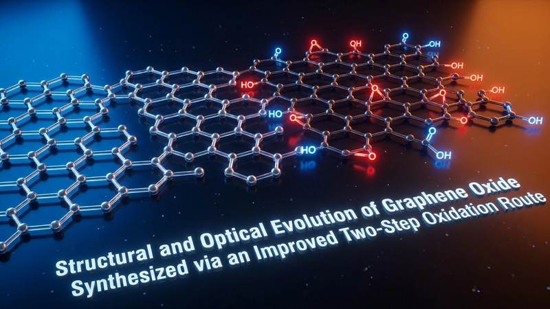
Understanding the fundamental properties of graphene oxide requires a comprehensive exploration of how its carbon lattice is altered during chemical processing. Unlike pristine graphene, which boasts a perfect hexagonal array of sp2 hybridized carbon atoms, graphene oxide is a complex, non-stoichiometric macromolecule. Its basal planes and edges are decorated with a myriad of oxygen-containing functional groups, such as hydroxyls, epoxides, carbonyls, and carboxyls. These groups disrupt the planar geometry of the native graphite, introducing sp3 hybridized defect sites that fundamentally alter the material's electrical, optical, and structural characteristics. The improved two-step oxidation route is specifically designed to control the introduction of these functional groups, ensuring a high degree of oxidation while preserving the structural integrity of the underlying carbon framework to the greatest extent possible.
The Evolution of Graphene Oxide Synthesis
The history of graphene oxide synthesis is a fascinating narrative of chemical refinement and optimization. The earliest attempts to oxidize graphite date back to the mid-nineteenth century, with the pioneering work of Benjamin Brodie, who utilized a hazardous mixture of potassium chlorate and fuming nitric acid. While successful in producing what he termed graphitic acid, the process was incredibly dangerous, time-consuming, and prone to explosive reactions. Decades later, Staudenmaier improved upon this by adding concentrated sulfuric acid to the mix, although the safety concerns remained paramount.
It was not until the mid-twentieth century that William Hummers and Richard Offeman introduced a significantly safer and more efficient protocol, which came to be known as the Hummers method. This technique utilized a water-free mixture of concentrated sulfuric acid, sodium nitrate, and potassium permanganate to oxidize graphite in a matter of hours rather than days. The Hummers method quickly became the industry standard, providing a reliable pathway for producing bulk quantities of graphene oxide. However, as the demand for high-quality graphene derivatives surged in the twenty-first century, researchers began to identify critical shortcomings in the traditional Hummers approach, including the generation of toxic nitrogen dioxide gas from the sodium nitrate and the incomplete oxidation of larger graphite flakes.
To address these challenges, the scientific community has aggressively pursued modifications to the original protocol. The improved two-step oxidation route eliminates the use of sodium nitrate entirely, thereby completely eradicating the emission of toxic nitrogenous gases and rendering the process significantly more environmentally friendly. Furthermore, by dividing the oxidation process into distinct pre-oxidation and deep oxidation phases, researchers can achieve a much higher degree of control over the exothermic reactions involved. This carefully managed thermal profile prevents the localized overheating that often leads to excessive defect formation and the complete breakdown of the carbon lattice, resulting in a superior graphene oxide product with highly tunable properties.
Decoding the Improved Two-Step Oxidation Route
The chemical mechanics underlying the improved two-step oxidation route are both elegant and highly complex. The process typically begins with a pre-oxidation phase, where raw graphite powder is subjected to a relatively mild oxidative environment. This initial step often involves treating the graphite with a mixture of concentrated sulfuric acid and a milder oxidizing agent, or a carefully controlled small dose of potassium permanganate. The goal here is not to completely oxidize the material, but rather to intercalate the acid molecules between the tightly stacked graphene layers. This intercalation process expands the interlayer spacing of the graphite, creating what is known as expandable graphite. This expanded structure drastically increases the accessible surface area for the subsequent chemical reactions.
Once the pre-oxidation is complete, the material enters the second phase, characterized by deep, aggressive oxidation. The pre-treated, expanded graphite is introduced into a fresh mixture of concentrated sulfuric acid and a substantial excess of potassium permanganate. Because the graphite layers have already been forced apart during the first step, the active oxidizing species can easily penetrate deep into the bulk of the material. The potassium permanganate reacts with the sulfuric acid to form dimanganese heptoxide, an incredibly potent oxidizer that aggressively attacks the carbon double bonds. This reaction introduces hydroxyl and epoxide groups onto the basal planes of the carbon sheets, while carboxylic and carbonyl groups are predominantly formed at the edges of the flakes.
Temperature control is absolutely paramount during this second step. The reaction between dimanganese heptoxide and graphite is highly exothermic. If the temperature is allowed to spike uncontrolled, the oxidation can become overly aggressive, tearing the graphene sheets into tiny, useless fragments or completely combusting the carbon into carbon dioxide. The improved two-step method employs rigorous temperature management, often utilizing ice baths and slow, dropwise addition of reagents to maintain a stable thermal environment. Following the completion of the oxidation, the reaction is quenched with distilled water and hydrogen peroxide, the latter serving to reduce the unreacted potassium permanganate into soluble manganese sulfate. The final, and arguably most tedious, aspect of the synthesis is the extensive washing protocol. Repeated centrifugation and washing with dilute hydrochloric acid and deionized water are required to remove all residual metal ions and acids, culminating in a highly purified, viscous aqueous dispersion of graphene oxide.
X-Ray Diffraction Insights into Structural Metamorphosis
X-ray diffraction is arguably the most definitive analytical technique for tracking the structural metamorphosis of graphite into graphene oxide. By bouncing X-rays off the crystalline planes of the material and measuring the angles of diffraction, researchers can calculate the precise distance between the individual atomic layers using Bragg's Law. In its pristine form, natural flake graphite exhibits a highly intense, sharp diffraction peak known as the 002 peak, which typically appears at a diffraction angle of approximately twenty-six degrees. This specific angle corresponds to an interlayer spacing of roughly zero point three four nanometers, reflecting the tight, van der Waals-driven stacking of the unadulterated graphene sheets.
As the improved two-step oxidation process unfolds, the X-ray diffraction pattern undergoes a dramatic and highly characteristic transformation. The intense graphite peak at twenty-six degrees progressively diminishes and eventually disappears entirely, providing clear evidence that the original crystalline structure has been thoroughly disrupted. In its place, a new, broader diffraction peak emerges at a significantly lower angle, typically between ten and eleven degrees. This shift to a lower diffraction angle is the hallmark of successful graphene oxide synthesis. According to Bragg's Law, a lower diffraction angle corresponds to a larger interplanar distance. In the case of fully oxidized graphene oxide, the interlayer spacing expands to anywhere from zero point seven to zero point nine nanometers, more than double the spacing of raw graphite.
This massive expansion in the interlayer spacing is driven by two primary factors. First, the introduction of bulky oxygen-containing functional groups onto the basal planes of the carbon sheets acts as physical spacers, forcing the layers apart. Second, these highly polar functional groups attract and trap water molecules from the aqueous environment used during synthesis and washing. These intercalated water molecules form complex hydrogen-bonding networks between the graphene oxide sheets, further increasing the distance between them. The exact position of the graphene oxide peak in the X-ray diffraction pattern can even be used as a proxy for environmental humidity, as the material will dynamically absorb and release water, causing the layers to swell and contract. Furthermore, the broadening of the diffraction peak compared to the sharp graphite peak indicates a significant reduction in the crystallite size along the vertical axis, confirming that the bulk graphite has been successfully exfoliated into few-layer or single-layer graphene oxide sheets.
Ultraviolet-Visible Spectroscopy and Electronic Transitions
While X-ray diffraction provides a macroscopic view of the structural changes, ultraviolet-visible spectroscopy offers a window into the fundamental alterations of the material's electronic band structure. Pristine graphene is a zero-bandgap semimetal, characterized by a continuous, unbroken network of sp2 hybridized carbon atoms that allow electrons to move freely across the basal plane. This continuous pi-electron conjugation results in strong, relatively featureless absorption across the entire visible and ultraviolet spectrum, which is why graphite appears black. However, the aggressive chemical oxidation process fundamentally destroys this perfect conjugation.
The introduction of oxygen functional groups converts a significant portion of the sp2 hybridized carbon atoms into sp3 hybridized state. This transformation creates localized electronic states and opens up a discernible bandgap, transforming the material from a conductive semimetal into a semiconductor or even a near-insulator. Ultraviolet-visible spectroscopy captures this electronic evolution beautifully. A typical absorption spectrum of an aqueous graphene oxide dispersion exhibits two distinct, highly characteristic features. The most prominent is a strong absorption peak located at a wavelength of approximately two hundred and thirty nanometers. This peak is attributed to the pi to pi star electronic transitions of the remaining unoxidized carbon-carbon double bonds within the disrupted lattice.
The second characteristic feature is a distinct shoulder peak occurring at around three hundred nanometers. This shoulder is assigned to the n to pi star transitions of the non-bonding electrons present in the oxygen atoms of the carbonyl and carboxyl functional groups. The relative intensities and precise wavelength positions of these two peaks provide a wealth of information regarding the degree of oxidation. In the improved two-step synthesis route, the high efficiency of the deep oxidation phase is reflected in the intense, well-defined nature of these peaks. Furthermore, researchers can utilize the ultraviolet-visible absorption data to calculate the optical bandgap of the synthesized graphene oxide using Tauc plots. By tracking how the bandgap changes in response to thermal or chemical reduction treatments, scientists can finely tune the electronic properties of the material, tailoring it for specific applications in optoelectronics, photocatalysis, or transparent conductive films.
Morphological Mapping with FESEM and EDS
To truly appreciate the physical nature of graphene oxide, researchers turn to field emission scanning electron microscopy. Unlike traditional scanning electron microscopy, the field emission variant utilizes a highly focused electron beam generated by a field emission gun, allowing for significantly higher resolution imaging at lower accelerating voltages. This is particularly crucial for analyzing carbonaceous nanomaterials like graphene oxide, which can easily be damaged or distorted by high-energy electron beams. The images produced by this technique reveal a morphology that is vastly different from the rigid, blocky appearance of raw flake graphite.
The field emission scanning electron microscopy images of graphene oxide synthesized via the improved two-step route typically display a highly folded, wrinkled, and crumpled sheet-like architecture. These sheets often resemble crumpled tissue paper or silk veils draped over one another. This characteristic wrinkling is not an artifact of the imaging process but a fundamental thermodynamic property of the material. The introduction of sp3 hybridized defect sites and bulky oxygen functional groups disrupts the planar geometry of the native graphene lattice, causing the two-dimensional sheets to buckle and fold in order to minimize their surface energy. This highly textured morphology is incredibly advantageous for many applications, as it prevents the sheets from tightly restacking, thereby maintaining a massive electrochemically active surface area.
Complementing the visual data provided by the electron microscopy is energy-dispersive X-ray spectroscopy, a technique used to determine the elemental composition of the sample. By analyzing the X-rays emitted by the sample when bombarded with the electron beam, researchers can quantify the ratio of carbon to oxygen atoms. A successful improved two-step oxidation will typically yield an energy-dispersive X-ray spectroscopy profile dominated exclusively by carbon and oxygen peaks, with the carbon to oxygen atomic ratio often falling between two to one and three to one, indicating a heavy degree of oxidation. Crucially, this spectroscopy technique also serves as a quality control metric for the washing process. The absence of peaks corresponding to sulfur, manganese, or potassium confirms that the rigorous purification steps were successful in removing the harsh chemicals used during the synthesis, ensuring that the final graphene oxide product is highly pure and ready for downstream integration.
Implications for Future Nanomaterial Applications
The rigorous characterization of graphene oxide synthesized via the improved two-step oxidation route is not merely an academic exercise; it has profound implications for the commercial and industrial application of nanomaterials. By understanding exactly how the structural and optical properties evolve, engineers can begin to leverage this material in highly specialized technologies. One of the most promising avenues is in the realm of advanced energy storage. The wrinkled, high-surface-area morphology observed in the electron microscopy images, combined with the presence of oxygen functional groups, makes graphene oxide an excellent precursor for supercapacitor electrodes. When partially reduced, the material regains much of its electrical conductivity while retaining the porous architecture necessary for rapid ion transport and high capacitance.
Furthermore, the exact tuning of the interlayer spacing, as confirmed by X-ray diffraction, is revolutionizing the field of membrane separation and water purification. By carefully controlling the degree of oxidation and the subsequent assembly of the graphene oxide sheets, researchers can create highly selective filtration membranes. These membranes feature nano-capillary networks that are precisely sized to allow water molecules to pass through while completely blocking larger ions, heavy metals, and organic pollutants. This technology holds immense promise for developing highly efficient, low-energy desalination plants and industrial wastewater treatment facilities.
Beyond energy and environmental applications, the tunable optical bandgap revealed by ultraviolet-visible spectroscopy opens doors in the biomedical and sensor sectors. The fluorescent properties of nanoscale graphene oxide can be harnessed for targeted drug delivery tracking and cellular imaging. Additionally, the abundant oxygen functional groups provide ideal anchor points for attaching specific enzymes or antibodies, allowing the creation of highly sensitive electrochemical biosensors capable of detecting disease markers at incredibly low concentrations. The improved two-step synthesis method is the critical enabler for all these technologies, providing a scalable, reliable, and high-yield pathway to produce the premium quality graphene oxide required to drive the next generation of nanotechnological innovation.
Frequently Asked Questions
Question: What is the primary advantage of the modified two-step Hummers method over the traditional approach?
Answer: The primary advantage lies in the enhanced control over the oxidation process, which results in a more uniform distribution of oxygen functional groups and a higher yield of single-layer graphene oxide. By separating the oxidation into two distinct phases, researchers can mitigate the explosive risks associated with rapid temperature spikes and reduce the formation of unwanted permanent defects in the carbon lattice. Furthermore, the elimination of sodium nitrate makes the process significantly safer and more environmentally friendly by preventing the release of toxic nitrogen dioxide gas. This careful modulation ultimately produces a higher quality material that is easier to functionalize for specific industrial applications.
Question: How does X-ray diffraction confirm the successful formation of graphene oxide from raw graphite?
Answer: X-ray diffraction confirms the formation of graphene oxide by measuring the shift in the primary diffraction peak, which corresponds to the distance between the layers of the material. Raw graphite exhibits a sharp peak at roughly twenty-six degrees, indicating a tight interlayer spacing of zero point three four nanometers. Following successful oxidation, this peak disappears completely and is replaced by a broader peak at around ten to eleven degrees. This shift proves that the interlayer spacing has expanded to zero point seven nanometers or more, driven by the intercalation of bulky oxygen functional groups and water molecules, verifying that the crystalline structure of the graphite has been thoroughly exfoliated and oxidized.
Question: What role does ultraviolet-visible spectroscopy play in analyzing the electronic properties of graphene oxide?
Answer: Ultraviolet-visible spectroscopy is critical for understanding how the chemical oxidation process alters the electronic band structure of the material. By measuring how the material absorbs different wavelengths of light, researchers can track the disruption of the continuous carbon double-bond network. The spectrum typically reveals a main absorption peak at two hundred and thirty nanometers, representing the remaining carbon-carbon bonds, and a shoulder at three hundred nanometers, representing the newly formed oxygen groups. Analyzing these peaks allows scientists to calculate the optical bandgap, confirming the transformation of the material from a conductive semimetal into a semiconductor.
Question: Why is the morphology of graphene oxide consistently described as wrinkled or crumpled in electron microscopy imaging?
Answer: The wrinkled and crumpled morphology observed in field emission scanning electron microscopy is a direct thermodynamic consequence of the chemical oxidation process. Pristine graphene is a perfectly flat, two-dimensional sheet due to its uniform sp2 hybridized carbon lattice. However, the aggressive oxidation introduces sp3 hybridized defect sites and attaches large, bulky oxygen functional groups to the surface. These physical disruptions destroy the planar geometry of the sheet, causing it to fold, buckle, and crumple in order to achieve a lower, more stable energy state. This wrinkling is highly beneficial as it prevents the sheets from clumping tightly together, thereby preserving a massive surface area.
Question: How does the improved synthesis method impact the environmental footprint of producing graphene oxide?
Answer: The improved two-step synthesis method significantly reduces the environmental footprint of graphene oxide production primarily by eliminating the use of sodium nitrate, which was a staple in the traditional Hummers method. The absence of sodium nitrate means that highly toxic and corrosive nitrogen dioxide gas is no longer generated and released into the atmosphere during the reaction. Additionally, the enhanced efficiency of the two-step process often results in a higher yield of usable material, reducing the overall volume of chemical waste and the amount of water required during the extensive purification and washing stages.
Conclusion
The rigorous investigation into the structural and optical evolution of graphene oxide synthesized via an improved two-step oxidation route marks a significant milestone in materials science. Through the comprehensive analysis of X-ray diffraction patterns, ultraviolet-visible spectra, and high-resolution electron microscopy, researchers have painted a detailed picture of how raw graphite transforms into a highly functional, tunable nanomaterial. The shift away from traditional, more hazardous synthesis methods toward this refined, two-step approach not only yields a safer and more environmentally conscious production process but also results in a vastly superior product.
By carefully controlling the introduction of oxygen functional groups and managing the expansion of the carbon lattice, scientists can now produce graphene oxide with precise structural and electronic characteristics. This level of control is absolutely essential for the transition of graphene-based technologies from laboratory curiosities to commercial realities. Whether it is serving as the backbone for next-generation supercapacitors, acting as the ultimate selective barrier in water filtration membranes, or enabling highly sensitive biomedical diagnostics, the future of this extraordinary material is brighter than ever. The foundational data and insights provided by this research will undoubtedly serve as a crucial guidepost for engineers and chemists as they continue to push the boundaries of what is possible in the realm of nanotechnology.
Evaluate Our Quality
Serious about B2B integration? Test our premium Pulsed Electrical Resistive Carbon Heating turbostratic graphene in your lab. 100g sample packs available now.