A Deep Dive into the Structural and Optical Evolution of Graphene Oxide via a Two-Step Oxidation Route
Research conducted by: Amani Azaizia, Andrey Yu. Ivanov, Idrisa Kiryowa, M. V. Dorogov
This remarkable team of researchers has provided the scientific community with an invaluable dataset and comprehensive analysis detailing the structural and optical evolution of graphene oxide. Their rigorous investigation, utilizing a modified two-step Hummers method, sheds light on the complex transformations that graphite undergoes during heavy oxidation. By meticulously documenting these changes through advanced characterization techniques, they have established a robust framework for understanding how precise chemical modifications can tailor the electronic and morphological properties of two-dimensional nanomaterials. We at usa-graphene.com recognize this work as a cornerstone for future scaling and functionalization efforts in the global graphene industry, offering a blueprint for manufacturing high-quality, defect-engineered nanostructures.
The Imperative for an Improved Oxidation Route
The synthesis of graphene oxide has historically been a challenging endeavor, balancing the need for thorough oxidation against the risks of structural degradation and environmental toxicity. The traditional Hummers method, developed in the late 1950s, utilizes a potent mixture of potassium permanganate, sodium nitrate, and concentrated sulfuric acid to force oxygen-containing functional groups into the tightly bound lattice of natural graphite. While revolutionary for its time, this original method is fraught with inefficiencies. It often results in incomplete oxidation, leaving behind unreacted graphite cores shielded by oxidized outer shells. Furthermore, the generation of toxic nitrogen dioxide gas presents severe safety and environmental hazards that complicate industrial-scale production.
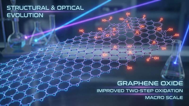
The improved two-step oxidation route analyzed in this research elegantly addresses these historical shortcomings. By breaking the oxidation process into distinct, carefully controlled phases, the researchers ensure a more uniform and pervasive functionalization of the carbon lattice. The first step acts as a pre-intercalation phase, gently expanding the interlayer spacing of the bulk graphite. This preliminary expansion acts as a gateway, allowing the primary oxidizing agents in the second step to penetrate deeply and uniformly throughout the entire graphite matrix. The elimination of sodium nitrate from the reaction mixture not only prevents the release of noxious gases but also refines the chemical environment, reducing unwanted byproducts. This two-step methodology represents a paradigm shift in nanomaterial synthesis, transitioning from a brute-force chemical assault to a highly tuned, sequential modification that preserves the foundational integrity of the two-dimensional sheets while maximizing their chemical reactivity.
X-Ray Diffraction and the Architecture of Interlayer Spacing
To truly understand the metamorphosis of graphite into graphene oxide, one must look at the atomic architecture, an endeavor made possible through X-ray diffraction. This analytical technique relies on the principles of constructive interference, governed by Bragg equation, to measure the distances between crystallographic planes. In pristine graphite, the carbon layers are held together by relatively weak van der Waals forces, resulting in a tightly packed structure with a characteristic interlayer spacing, or d-spacing, of approximately 0.34 nanometers. This manifests in an X-ray diffraction pattern as a sharp, highly intense peak at a specific diffraction angle.
The data provided by the research team reveals a dramatic structural evolution following the two-step oxidation process. As the potent chemical oxidants drive oxygen functional groups such as epoxides, hydroxyls, and carboxyls into the carbon framework, these bulky groups act as molecular wedges. They physically force the individual graphene sheets apart. Additionally, the hydrophilic nature of these newly introduced functional groups attracts water molecules from the aqueous reaction environment, which further intercalate between the layers.
Consequently, the X-ray diffraction pattern undergoes a profound transformation. The prominent graphite peak completely disappears, replaced by a new, broader peak at a significantly lower diffraction angle. Calculations derived from this new peak demonstrate that the interlayer spacing has expanded massively, often exceeding 0.80 nanometers. This more than doubling of the d-spacing is the definitive crystallographic signature of successful graphene oxide synthesis. The broadening of the peak also provides critical information about the crystallite size along the stacking axis. Using the Scherrer equation, scientists can deduce that the long-range order of the bulk graphite has been systematically dismantled, resulting in an exfoliated material comprising only a few, highly functionalized layers. This expanded architecture is precisely what makes graphene oxide dispersible in water and highly amenable to subsequent chemical functionalization for advanced industrial applications.
Ultraviolet-Visible Spectroscopy and Electronic Band Gap Tuning
The physical expansion of the crystal lattice is intimately tied to a fundamental restructuring of the material electronic properties, a phenomenon expertly captured through ultraviolet-visible spectroscopy. Pristine graphene is famous for its zero-bandgap electronic structure, where highly delocalized pi electrons move freely across the planar hexagonal lattice, granting the material its extraordinary electrical conductivity. However, the introduction of oxygen functional groups during the two-step oxidation process violently disrupts this conjugated network. The sp2 hybridized carbon atoms, which form the flat, conductive sheets, are converted into sp3 hybridized atoms wherever an oxygen group attaches. This transition forces the electrons into localized bonds, effectively opening an electronic bandgap and transforming the conductive graphene into an insulating or semiconducting graphene oxide.
The ultraviolet-visible absorption spectra documented in this research provide a clear window into this electronic evolution. The spectrum of the synthesized graphene oxide typically features two distinct absorption characteristics. The first is a prominent maximum absorption peak located around 230 nanometers. This peak corresponds to the pi to pi star electronic transitions occurring within the remaining, unoxidized islands of sp2 carbon-carbon bonds. The intensity and exact wavelength of this peak serve as a reliable metric for the extent of the surviving conjugated network.
The second defining feature is a distinct shoulder peak emerging around 300 nanometers. This secondary absorption event is attributed to the n to pi star transitions associated with the carbon-oxygen double bonds of the carbonyl and carboxyl functional groups that decorate the edges and basal planes of the sheets. By monitoring the shifts and intensities of these peaks, researchers can precisely quantify the degree of oxidation achieved by the two-step method. Furthermore, by applying Tauc plot methodologies to the absorption data, scientists can calculate the exact optical bandgap of the material. This capability to tune the bandgap through controlled oxidation is incredibly powerful, as it allows engineers to customize the optoelectronic properties of the graphene oxide for specific applications, ranging from transparent conductive films to active layers in photovoltaic cells.
Morphological Insights from Field Emission Scanning Electron Microscopy
While crystallographic and spectroscopic data provide essential information about the atomic and electronic states of the material, understanding its macroscopic physical behavior requires direct visual observation. Field emission scanning electron microscopy offers the nanometer-scale resolution necessary to observe the morphological evolution of the oxidized sheets. Unlike standard electron microscopes, field emission variants utilize a highly focused, low-energy electron beam that minimizes charging effects and sample damage, providing exceptionally crisp, high-contrast images of delicate two-dimensional structures.
The images generated in this study reveal that the idealized concept of perfectly flat graphene sheets does not apply to heavily oxidized graphene oxide. Instead, the field emission scanning electron microscopy images showcase a highly complex, chaotic topography characterized by extensive wrinkling, folding, and corrugation. The material often resembles crumpled silk or heavily textured paper. This wrinkled morphology is not a defect of the synthesis process but rather a fundamental physical consequence of the chemical oxidation.
As previously noted, the attachment of oxygen functional groups converts planar sp2 carbon atoms into tetrahedral sp3 carbon atoms. This localized change in geometry introduces immense structural strain into the two-dimensional lattice. The sheet buckles and folds to relieve this thermodynamic stress, resulting in the wrinkled appearance observed under the microscope. This unique morphology is highly advantageous for many commercial applications. The wrinkles and folds act as physical spacers, preventing the individual graphene oxide sheets from tightly restacking via van der Waals interactions when they are dried or integrated into solid-state devices. By inhibiting restacking, the wrinkled morphology preserves a massive electrochemically active surface area, which is an absolute necessity for the development of high-capacity supercapacitors, advanced battery electrodes, and highly sensitive chemical sensors.
Energy Dispersive Spectroscopy and Elemental Mapping
To corroborate the structural and morphological findings, the researchers employed energy dispersive spectroscopy in tandem with the electron microscopy. This analytical technique analyzes the X-rays emitted by the sample when bombarded by the electron beam, providing a precise, localized quantification of the elemental composition. For graphene oxide synthesized via the improved two-step route, this data is critical for confirming the efficiency and uniformity of the oxidation process.
The energy dispersive spectroscopy data yields the crucial carbon-to-oxygen atomic ratio. In pristine graphite, this ratio is effectively infinite, as oxygen is entirely absent from the pure carbon lattice. Following the rigorous two-step oxidation, the data typically reveals a carbon-to-oxygen ratio approaching two to one, or sometimes even lower depending on the precise reaction parameters. This high concentration of oxygen confirms that the two-step method is exceptionally efficient at driving functional groups into the material framework.
Equally important is the spatial distribution of these elements. Advanced elemental mapping techniques demonstrate that the oxygen is distributed highly uniformly across the entire scanned area of the graphene oxide flakes. This homogeneity is a direct triumph of the two-step method pre-intercalation phase, which ensures that the oxidants do not merely react with the outer edges of the graphite particles but penetrate deeply to functionalize the core. The spectroscopy data also serves a vital quality control function by detecting trace impurities. By monitoring the levels of sulfur or manganese remaining from the sulfuric acid and potassium permanganate reagents, researchers can evaluate the effectiveness of their post-synthesis washing and purification protocols, ensuring that the final graphene oxide product meets the stringent purity standards required for advanced electronic and biomedical applications.
Implications for Advanced Optoelectronic and Energy Devices
The exhaustive characterization provided by this research extends far beyond basic material science; it lays the foundation for the next generation of advanced technologies. The structural and optical evolution documented here directly dictates how this material can be utilized in the commercial sector. The ability to finely tune the interlayer spacing, as proven by the X-ray diffraction data, makes this specific type of graphene oxide an ideal candidate for advanced filtration membranes. By controlling the d-spacing, engineers can create selective sieves capable of desalinating seawater or filtering heavy metal toxins with unprecedented efficiency and throughput.
Furthermore, the electronic bandgap tuning revealed by the ultraviolet-visible spectroscopy opens massive opportunities in optoelectronics. Because the oxidation process allows the material to transition from a conductor to a semiconductor, carefully reduced versions of this graphene oxide can be used to manufacture flexible, transparent conductive films for touchscreens and organic light-emitting diode displays. The tunable bandgap also makes it a highly attractive material for photocatalysis and solar energy harvesting, where matching the absorption spectrum to the solar emission spectrum is critical for maximizing energy conversion efficiency.
Finally, the highly wrinkled morphology and massive surface area confirmed by the field emission scanning electron microscopy are perfectly suited for energy storage applications. When utilized as an electrode material in supercapacitors or lithium-ion batteries, the uniform distribution of oxygen groups provides excellent wettability with aqueous and organic electrolytes. The structural wrinkles prevent the sheets from aggregating, ensuring that ions have unimpeded access to the entire surface area of the electrode. This translates to faster charging times, higher energy densities, and longer operational lifespans for consumer electronics and electric vehicles. The research conducted by this team thus provides a vital roadmap for translating raw graphite into high-performance, commercially viable energy solutions.
Frequently Asked Questions
QUESTION: What is the primary advantage of the modified two-step oxidation route over the traditional method?
ANSWER: The modified two-step route offers significant improvements in both reaction efficiency and safety. By utilizing a pre-oxidation step to gently expand the graphite lattice, the primary oxidants can penetrate the material much more thoroughly, resulting in a highly uniform distribution of oxygen functional groups. Additionally, by removing sodium nitrate from the chemical formulation, the modified method eliminates the release of toxic nitrogen dioxide gas, making the process substantially safer for laboratory personnel and much more viable for large-scale industrial manufacturing.
QUESTION: How does X-ray diffraction confirm the successful synthesis of graphene oxide?
ANSWER: X-ray diffraction measures the distance between the atomic layers in a crystal. Pristine graphite has a very tight interlayer spacing, showing a sharp peak at a specific angle. When graphite is successfully oxidized into graphene oxide, bulky oxygen functional groups and water molecules force the carbon layers apart. This causes the original graphite peak to disappear and a new peak to emerge at a lower angle, indicating that the interlayer spacing has more than doubled. This massive expansion is the definitive proof of successful oxidation.
QUESTION: Why does graphene oxide appear wrinkled and folded under an electron microscope?
ANSWER: The wrinkling is a physical response to the chemical changes occurring at the atomic level. During oxidation, planar sp2 hybridized carbon atoms are converted into tetrahedral sp3 hybridized atoms as oxygen groups attach to the lattice. This change in geometric structure introduces severe mechanical strain into the previously flat two-dimensional sheet. To relieve this thermodynamic stress, the sheet buckles, folds, and wrinkles. This corrugated morphology is actually highly beneficial, as it prevents the sheets from clumping together and preserves a high surface area for chemical reactions.
QUESTION: What role does ultraviolet-visible spectroscopy play in analyzing these materials?
ANSWER: Ultraviolet-visible spectroscopy measures how the material absorbs light at different wavelengths, which provides direct insight into its electronic structure. The spectrum of graphene oxide shows specific absorption peaks that correspond to the electronic transitions of carbon-carbon and carbon-oxygen bonds. By analyzing the position and intensity of these peaks, scientists can determine the extent of the conjugated carbon network that remains intact and calculate the optical bandgap of the material. This tells researchers whether the material will behave as a conductor, semiconductor, or insulator.
QUESTION: Can this modified synthesis method be scaled up for commercial industrial production?
ANSWER: Yes, the modified two-step method is highly amenable to industrial scale-up. The elimination of toxic gas byproducts simplifies the necessary environmental controls and safety infrastructure required for a manufacturing plant. Furthermore, the enhanced uniformity of the oxidation process ensures a more consistent, higher-yield product, reducing batch-to-batch variability. As the demand for high-quality graphene oxide increases in sectors like energy storage, water filtration, and optoelectronics, this safer and more efficient synthetic route will play a critical role in meeting global supply requirements.
Conclusion
The comprehensive dataset and analysis provided by this research team represent a significant leap forward in our understanding of two-dimensional nanomaterials. By meticulously tracking the structural and optical evolution of graphene oxide synthesized via an improved two-step route, they have demystified the complex physical and chemical transformations that dictate material performance. The integration of advanced X-ray diffraction, ultraviolet-visible spectroscopy, and electron microscopy offers a masterclass in nanomaterial characterization. As the global industry continues to push the boundaries of what is possible with advanced carbon materials, the insights gleaned from this study will undoubtedly accelerate the development of next-generation energy storage devices, flexible electronics, and advanced filtration systems. The transition from theoretical potential to commercial reality relies entirely on this level of rigorous, foundational science.
Evaluate Our Quality
Serious about B2B integration? Test our premium Pulsed Electrical Resistive Carbon Heating turbostratic graphene in your lab. 100g sample packs available now.